
BalCare — чат


Dental Blog

Read our dental blog to learn more about your oral health, get tips on dental care, and discover helpful articles about your smile. We take pride in creating a friendly, positive dental environment where you can always feel comfortable under our care and guidance.

miofascialna-bolka-glavata-dentalna-klinika-balcare.webp
27/Apr/2026

Myofascial Head Pain: The Hidden Cause of Tension, Headaches and Disturbed Sleep – and How Red Light Therapy May Help

Myofascial pain in the head and facial region is a functional disorder that often remains undiagnosed, despite having a significant impact on patients’ quality of life. It can present with a wide range of symptoms – from diffuse facial pain and headaches to a feeling of tension, discomfort while chewing, and disturbed sleep.

In the clinical practice of BalCare Dental Clinic, these complaints are assessed in the context of myofascial pain syndrome, in which the masticatory muscles, fascial structures, and neural regulation function under conditions of chronic tension.

Not every type of head pain has the same origin, even though different conditions may feel very similar to the patient. In clinical practice, it is essential to distinguish between muscular (myofascial) pain and joint-related pain, because this determines both the underlying cause and the appropriate treatment approach.

In myofascial pain, the primary problem lies within the masticatory muscles. The muscles remain in a state of chronic tension or spasm, often as a result of teeth clenching, bruxism, or prolonged stress-related loading.

This type of pain is usually not clearly localised. It may be experienced as deep, diffuse tension and can radiate towards the temple, ear, or teeth. It is often more pronounced in the morning, when the patient wakes with a feeling of stiffness or muscular fatigue.

By contrast, joint disorders affect the intra-articular structures of the temporomandibular joint (TMJ).

In these cases, the pain tends to be more clearly localised in front of the ear and is usually triggered or intensified by movement, such as wide mouth opening or chewing.

It may also be accompanied by clicking, popping, or restricted movement, suggesting a mechanical problem within the joint itself.

This distinction has direct practical importance.

When the source of pain is muscular, the therapeutic focus is on reducing muscle tone and restoring normal function. It is in these cases that red light therapy (photobiomodulation) may be particularly beneficial, as it can support muscle relaxation, improve local circulation, and help reduce inflammatory processes.

In joint-related disorders, however, a more comprehensive approach may be required, focusing on the structures within the joint itself.

For this reason, accurate diagnosis is essential. It allows the clinician to determine not only where the pain originates, but also which therapeutic approach may be most appropriate for the individual patient.

Pathophysiology of Myofascial Pain

Myofascial pain develops as a result of prolonged muscular overload, which can lead to the formation of trigger points – localised areas of increased muscle tone and altered sensitivity.

A characteristic feature of these points is their ability to cause referred pain, meaning that pain is perceived in an area different from the actual source.

This can create diagnostic difficulties and may lead patients to consult different medical specialists without the underlying cause being identified.

Clinical Importance of the Masseter and Temporalis Muscles

The muscles most commonly involved in temporomandibular myofascial pain are the masseter and temporalis muscles, because of their central role in chewing and their tendency to become overloaded in patients with bruxism.

When trigger points are present in the masseter muscle, referred pain may be felt in the lower jaw, posterior teeth, and the area around the ear.

Patients often report increased tooth sensitivity without any objective dental findings, as well as discomfort during chewing and morning jaw stiffness.

The temporalis muscle, on the other hand, is associated with pain localised in the temple and forehead.

These symptoms are frequently interpreted as tension-type headaches without recognition of their possible muscular origin.

Trigger Points in the Masseter and Temporalis – A Common Cause of Referred Facial Pain

In clinical practice, the masseter and temporalis muscles are among the most common sources of myofascial pain in the head and facial region.

They are the primary muscles involved in closing the mouth and play an important role in clenching and bruxism.

When chronically overloaded, these muscles may develop trigger points – localised areas of increased muscle tone that can cause referred pain in different areas of the face and head.

This means that the actual source of the pain and the area where the pain is felt often do not coincide.

This mechanism helps explain why patients may report symptoms that cannot be linked to a specific structural pathology.

Masseter Muscle – Referred Pain to the Teeth, Jaw and Ear

The masseter is one of the principal muscles of mastication and is exposed to significant functional loading, particularly in patients with bruxism.

When active trigger points are present in the masseter, the following symptoms may occur:

  • pain in the lower jaw;
  • increased sensitivity in the posterior teeth without a dental cause;
  • discomfort while chewing;
  • tension and stiffness in the jaw, particularly in the morning;
  • referred pain towards the ear.

These symptoms often require differential diagnosis from odontogenic and ear, nose and throat conditions.

Temporalis Muscle – Referred Pain to the Temple and Forehead

The temporalis muscle plays an active role in stabilising the lower jaw and is particularly sensitive to chronic tension.

Trigger points in this muscle may cause:

  • pain in the temple region;
  • pain spreading towards the forehead;
  • a sensation of diffuse tension in the upper part of the face;
  • headache characterised by prolonged pressure.

This type of pain is often interpreted as a tension-type headache without consideration of its muscular origin.

Symptom Location of Pain Likely Muscular Source Clinical Characteristics
Pain in the posterior teeth without objective findings Lower jaw Masseter Intensifies during clenching or chewing
Morning stiffness and tension Jaw, cheeks Masseter Associated with nocturnal bruxism
Pain around the ear Periauricular area Masseter / pterygoid muscles Without evidence of ear pathology
Headache with diffuse pressure Temple, forehead Temporalis muscle Persistent, often bilateral
Pain during wide mouth opening Temporomandibular joint region Lateral pterygoid Intensifies with movement
Combined neck and facial pain Back of the head, jaw SCM / trapezius Associated with posture and muscular imbalance

The Relationship Between Myofascial Pain and Sleep

Chronic muscle tension may directly affect sleep quality.

Increased muscle tone and persistent pain can disturb normal sleep architecture, including through frequent micro-awakenings and a reduction in the deeper stages of sleep.

Patients often report:

  • non-restorative sleep;
  • morning fatigue;
  • headache on waking;
  • a feeling of tension in the jaw.

Scientific analyses have reported statistically significant reductions in sleep quality among patients with temporomandibular myofascial pain.

The Role of Bruxism and Stress

Myofascial pain is often associated with bruxism – involuntary clenching or grinding of the teeth, frequently occurring during sleep.

This process increases muscular loading and can contribute to the persistence of chronic pain.

Stress also plays an important role as an aggravating factor because it can increase muscle tone and activate neuromuscular mechanisms that make relaxation more difficult, even at rest.

Red Light Therapy (Photobiomodulation)

Photobiomodulation is a non-invasive therapeutic method that uses light of a specific wavelength to influence cellular processes.

Its proposed mechanisms of action include:

  • stimulation of mitochondrial activity;
  • increased ATP production;
  • improvement of local microcirculation;
  • reduction of inflammatory mediators.

As a result, pain may be reduced and muscular function may improve.

Clinical Approach at BalCare Dental Clinic

At BalCare Dental Clinic, red light therapy is used as part of a structured gnathological approach that includes functional diagnostics, assessment of muscle balance, and an individualised treatment plan.

In acute conditions, therapy is directed towards reducing pain and muscle spasm.

In chronic cases, the aim is to restore muscular function and stabilise the condition.

It is important to emphasise that photobiomodulation is not used as an isolated procedure, but as part of a comprehensive treatment approach that may include other therapeutic methods depending on the individual case.

Additional information about red light therapy:
https://balcare.bg/red-light-therapy/

Myofascial pain in the head and facial region is a complex condition that affects not only the local musculature but also sleep quality, neural regulation, and the patient’s everyday comfort.

Recognition of trigger points and accurate differential diagnosis are essential for effective treatment.

Modern therapeutic methods, including photobiomodulation, offer the possibility of targeting key pain mechanisms when applied according to an appropriate protocol and as part of a comprehensive treatment strategy.

Bibliography

https://pmc.ncbi.nlm.nih.gov/articles/PMC8291152/
Systematic review and meta-analysis on the effectiveness of photobiomodulation for myofascial pain associated with temporomandibular disorders.

https://pubmed.ncbi.nlm.nih.gov/29697718/
Systematic review of low-level light therapy for temporomandibular myofascial pain and its relationship with symptoms.

https://link.springer.com/article/10.1186/s12903-023-02784-8
Randomised clinical trial comparing the effects of LED and laser therapy in patients with temporomandibular disorders.

https://pmc.ncbi.nlm.nih.gov/articles/PMC7356229/
Review article on the cellular mechanisms of photobiomodulation.

temporomandibularna-stava-syn-denatalna-klinika-balcare.webp
22/Apr/2026

How Does the Temporomandibular Joint Affect Sleep?

Problems involving the temporomandibular joint (TMJ) are often viewed simply as local discomfort in the jaw area. In clinical practice, however, something much more significant is increasingly being observed – a direct relationship between temporomandibular disorders, chronic pain, and sleep disturbances.

Patients rarely associate morning fatigue, frequent awakenings, or nighttime teeth clenching with a problem involving the jaw joint, which may delay seeking appropriate treatment.

The temporomandibular joint is a small but extremely important structure that connects the lower jaw to the skull. It is involved every time we open the mouth, chew, speak, yawn, or swallow.

When this joint or the muscles surrounding it do not function properly, the problem rarely remains limited to “jaw pain”. Many patients begin to experience morning headaches, facial tension, pain around the ear, tooth sensitivity, stiffness when opening the mouth, and disturbed sleep.

At BalCare Dental Clinic, temporomandibular dysfunction is approached as a functional problem that may affect not only the bite and the masticatory muscles, but also sleep quality.

This is particularly important for patients who wake feeling tired, clench their teeth during the night, or feel that they are “unable to relax their jaw”.

Why Can a Jaw Problem Disturb Sleep?

Many patients do not associate poor sleep with their teeth or jaw.

They may look for the cause in stress, exhaustion, hormonal changes, an uncomfortable pillow, or everyday tension.

These factors may indeed contribute, but temporomandibular dysfunction introduces another important mechanism: the body may be unable to relax fully because the masticatory muscles remain active during the night.

When the masseter, temporalis, and other muscles surrounding the joint remain tense, the brain continues to receive signals of discomfort.

This may contribute to micro-awakenings that the patient does not remember the following morning but experiences indirectly as fatigue, irritability, and a lack of restorative sleep.

The Most Common Symptoms Patients Tend to Ignore

Temporomandibular dysfunction does not always begin with acute pain.

The first signs are often much more subtle:

  • morning jaw tension;
  • facial pain or fatigue;
  • headaches after waking;
  • clicking or popping when opening the mouth;
  • difficulty chewing;
  • pain around the ear without an identifiable ear-related cause;
  • teeth clenching or grinding;
  • tooth sensitivity;
  • disturbed, light, or fragmented sleep.

These symptoms are often treated separately – with painkillers, massage, a new pillow, or sleep supplements.

However, when the underlying cause involves the masticatory system, the relief is often only temporary.

Bruxism: Nighttime Clenching That Can Maintain the Problem

Bruxism is one of the important factors in patients with temporomandibular disorders and disturbed sleep.

It involves clenching or grinding of the teeth, often occurring unconsciously during sleep. This places additional load on the temporomandibular joint, masticatory muscles, and teeth.

The problem is that bruxism may create a vicious cycle.

The more a patient clenches their teeth, the more the muscles become overloaded. The greater the muscular tension, the more pronounced the pain may become. And the stronger the discomfort, the more difficult it may be for the body to reach deep, restorative sleep.

An important professional distinction should be made here: not every case of bruxism indicates severe temporomandibular dysfunction.

However, when bruxism is accompanied by pain, morning stiffness, headaches, and disturbed sleep, a gnathological assessment is recommended.

The Relationship Between TMD and Sleep Apnoea – What Patients Should Know

Sleep apnoea is a condition in which breathing repeatedly stops or becomes significantly reduced during sleep.

The most common form is obstructive sleep apnoea (OSA), in which the upper airway becomes narrowed or temporarily blocked.

This may lead to:

  • snoring;
  • frequent awakenings;
  • dry mouth in the morning;
  • daytime sleepiness;
  • headaches;
  • increased cardiovascular risk.

The relationship between temporomandibular disorders and sleep apnoea is complex and should not be oversimplified.

It would not be accurate to claim that TMD “causes” sleep apnoea in every patient.

A more precise explanation is that some patients may have overlapping factors, including:

  • the position of the lower jaw;
  • muscular tension;
  • bruxism;
  • disturbed breathing during sleep;
  • unstable or fragmented sleep.

More recent studies and review publications have reported an association between painful temporomandibular disorders and obstructive sleep apnoea, although the scientific evidence still requires careful interpretation.

A 2021 review indicated that the evidence supporting an association between TMD and sleep quality is stronger, while the relationship between TMD and sleep apnoea requires further high-quality research.

A more recent meta-analysis published in 2024 reported a significant association between TMD and obstructive sleep apnoea.

The practical message for patients is clear:

If jaw pain is accompanied by loud snoring, pauses in breathing, sudden awakenings with shortness of breath, excessive daytime sleepiness, or high blood pressure, consultation with a sleep medicine specialist or an ear, nose and throat (ENT) specialist is also necessary.

The role of the dentist and gnathologist is to assess jaw function, the bite, muscular tension, and the possible need for dental appliances.

However, sleep apnoea requires appropriate medical diagnosis.

How Does Red Light Therapy Fit Into Treatment?

Red light therapy, also known as photobiomodulation, is a modern non-invasive treatment method that uses red and/or near-infrared light with specific therapeutic parameters.

Its purpose is not simply to “warm” the area or provide temporary relaxation.

Instead, the therapy is intended to influence cellular processes, local circulation, inflammation, and tissue recovery.

In patients with temporomandibular dysfunction, therapy may be directed towards the joint area and the masticatory muscles when indicated after examination.

At BalCare Dental Clinic, photobiomodulation is not applied randomly or according to the principle that “more is better”.

Treatment follows specific protocols, with defined treatment areas, duration, and sequence of procedures according to the patient’s individual condition.

This distinction is important because photobiomodulation is a dose-dependent therapy.

Its effects depend on appropriate assessment, the correct area of application, and the correct treatment protocol.

A 2023 systematic review on photobiomodulation for temporomandibular disorders reported promising effects in reducing pain and improving maximum mouth opening.

How Can Reducing Pain Improve Sleep?

Red light therapy is not a sleeping medication.

It does not act as a drug for insomnia and should not be presented in this way.

Its potential value for patients with temporomandibular disorders lies in helping to reduce some of the factors that may interfere with sleep:

  • pain;
  • inflammation;
  • muscular tension.

When the masticatory muscles become more relaxed, it may be easier for the patient to reach a state of rest.

When pain is reduced, there may be fewer pain-related micro-awakenings.

When the joint is less irritated, morning stiffness and discomfort may also decrease.

This is the practical relationship between TMD, red light therapy, and sleep:

The treatment does not directly target “insomnia”; instead, it may help address one of the potential sources of nighttime discomfort.

The Approach at BalCare Dental Clinic

At BalCare Dental Clinic, red light therapy is considered part of a comprehensive treatment strategy rather than a universal stand-alone solution.

For patients experiencing jaw pain, bruxism, and disturbed sleep, the first step is to assess the function of the masticatory system.

The treatment plan may include:

  • gnathological assessment;
  • bite evaluation;
  • examination of the masticatory muscles;
  • assessment of the temporomandibular joint;
  • red light therapy according to an individual protocol;
  • a custom-made occlusal splint for bruxism when indicated;
  • guidance for reducing muscular overload;
  • follow-up assessment of treatment results.

What Is Important Not to Overlook?

One of the most important aspects often missing from discussions of this subject is the distinction between a symptom and its underlying cause.

Jaw pain may result from:

  • muscular overload;
  • abnormal functional loading;
  • bruxism;
  • a joint disorder;
  • stress;
  • sleep disturbances;
  • or a combination of several factors.

For this reason, treatment must be individualised.

In one patient, the primary problem may be nighttime clenching.

In another, it may be inflammation and pain within the joint.

A third patient may have sleep apnoea, in which case the dental approach needs to be coordinated with appropriate medical diagnosis.

For another patient, an unstable bite and chronic overloading of specific muscles may be the main contributing factors.

The professional approach is not to promise a quick universal solution, but to identify the precise mechanism behind the patient’s symptoms.

When Should You Seek a Consultation?

A professional assessment is recommended if you experience:

  • jaw pain in the morning;
  • teeth clenching or grinding;
  • frequent headaches;
  • clicking or popping in the joint;
  • facial tension;
  • pain around the ear;
  • fragmented sleep;
  • fatigue despite apparently sufficient hours of sleep;
  • snoring or suspected sleep apnoea.

When such symptoms are present, assessment should not be unnecessarily delayed.

The earlier the underlying cause is identified, the easier it may be to interrupt the cycle of:

pain – muscle tension – bruxism – disturbed sleep.

The temporomandibular joint may have a greater relationship with sleep quality than many patients realise.

When the jaw remains tense, the muscles are overloaded, and the joint is irritated, the resulting discomfort may make restorative sleep more difficult.

Scientific evidence supports an association between temporomandibular disorders and impaired sleep quality.

Photobiomodulation, or red light therapy, is a modern therapeutic method that may support the reduction of pain and muscular tension when it is appropriately indicated and applied according to a defined protocol.

At BalCare Dental Clinic, the approach is based on comprehensive assessment of the masticatory system, an individual treatment plan, and monitoring of the results.

The goal is not simply temporary symptom relief, but the restoration of function, comfort, and quality of life.


kakvo-predstavlyava-red-light-therapy-dentalna-klinika-balcare.webp
06/Apr/2026

What Is Red Light Therapy?

How to Prepare for Your First Session at BalCare Dental Clinic

What to expect: protocols, duration, sensations, and realistic results

At BalCare Dental Clinic, Red Light Therapy is used as a modern, gentle, and effective method for relieving jaw pain, bruxism, and muscular tension.

Many patients want to know what happens during the first session and what they can realistically expect from the therapy.

This article provides clear and practical information to help you prepare with confidence and get the maximum benefit from the treatment.

Stimulating Cellular Activity

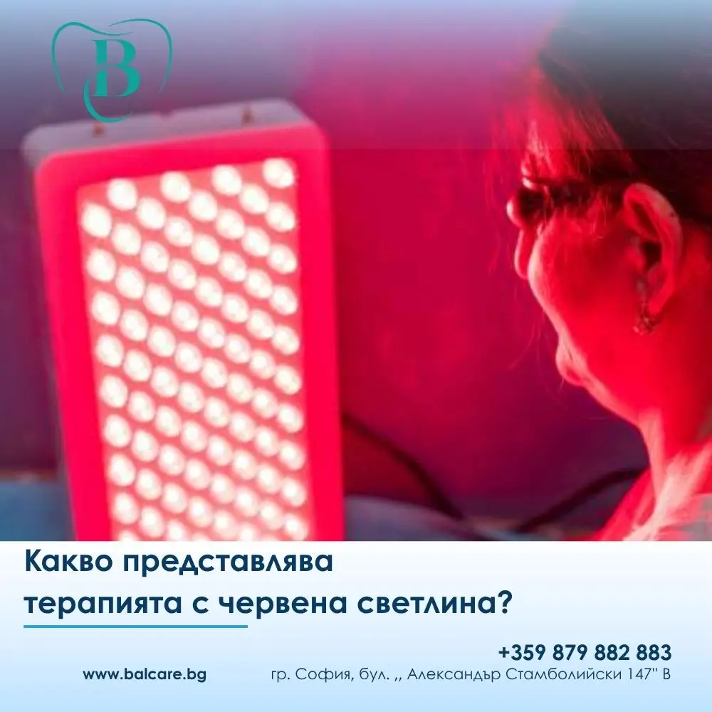
Red Light Therapy, also known as photobiomodulation, uses specific wavelengths of red and near-infrared light that penetrate the tissues and stimulate cellular activity.

This may contribute to:

  • reducing inflammation;
  • supporting tissue recovery;
  • relaxing the muscles;
  • reducing pain.

In dentistry, the method is used for conditions such as:

  • jaw pain;
  • temporomandibular dysfunction;
  • bruxism and teeth clenching;
  • muscular tension.

If you experience similar symptoms, you can also read our detailed article about jaw pain.

How to Prepare for Your First Session

Preparation is simple, but it can be important for the effectiveness of the therapy.

It is recommended that:

✔️ no creams or make-up are applied to the facial treatment area;
✔️ you inform the clinician about any medications you are taking and any relevant medical conditions;
✔️ you mention if you use an occlusal splint for bruxism.

No fasting or special preparation is required.

It is best to arrive as relaxed as possible, as stress can influence muscle tone.

What Happens During the First Appointment – Step by Step

The first appointment begins with a clinical assessment of your condition.

The clinician evaluates:

✔️ muscular tension;
✔️ painful or sensitive points;
✔️ range of motion;
✔️ temporomandibular joint function.

An individual treatment protocol is then prepared according to the specific problem.

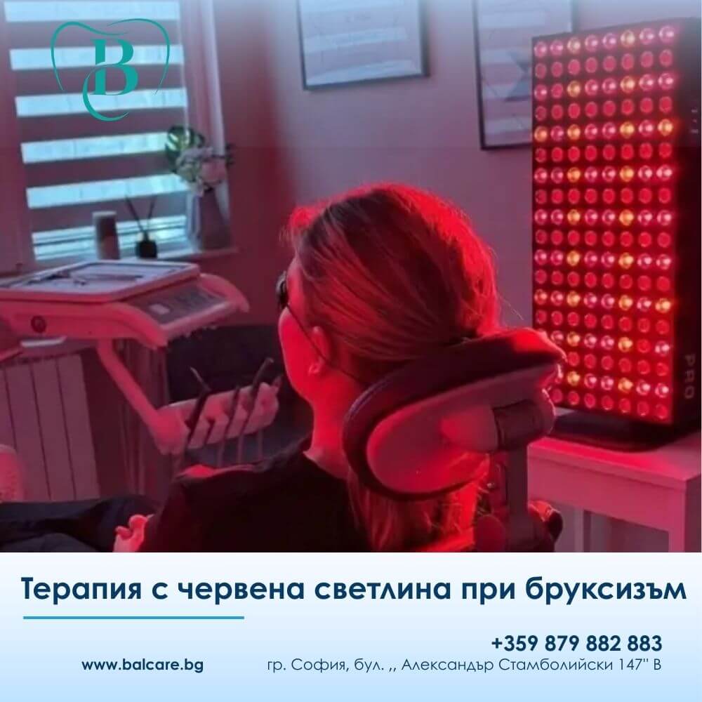
The patient is positioned comfortably, protective glasses are provided, and the light device is activated.

How Long Does the First Session Take?

The procedure usually lasts approximately 10 to 12 minutes per side.

For milder conditions, treatment may be shorter, while chronic conditions may require longer treatment and application to several areas.

What Will You Feel During the Therapy?

The therapy is completely painless.

The most common sensations include:

✔️ gentle warmth;
✔️ relaxation of the muscles;
✔️ an overall feeling of relaxation.

Some patients notice a reduction in tension even during the first treatment session.

What Results Can You Expect After the First Session?

After the first procedure, some patients may experience:

✔️ reduced pain;
✔️ less tension in the jaw;
✔️ greater comfort while chewing.

With consecutive sessions, the effect may gradually build and stabilise.

If you experience nighttime teeth clenching, you can also read: Red Light Therapy for Bruxism.

How Many Sessions Are Needed?

✔️ 12 sessions for muscular problems
✔️ 24 sessions for joint-related problems

Is Red Light Therapy Safe?

Yes, the therapy is safe and gentle when applied correctly.

✔️ No medication is used.
✔️ The tissues are not damaged.
✔️ When properly applied, the treatment is designed to minimise unwanted effects.

Are There Any Contraindications?

In certain situations, prior consultation is necessary, including:

✔️ photosensitivity;
✔️ use of certain medications;
✔️ oncological conditions;
✔️ pregnancy.

When Is Red Light Therapy Not Enough on Its Own?

Some conditions require a combined treatment approach.

Red Light Therapy may be combined with:

  • an occlusal splint for bruxism;
  • orthodontic treatment;
  • bite correction.

Can It Be Combined with Other Treatments?

Yes.

Red Light Therapy can be combined with different dental and restorative treatments and may support the recovery process.

What Should You NOT Do After the Procedure?

There are no specific restrictions required after the treatment.

Comparison with Other Methods

  • Medication – temporary symptom relief;
  • occlusal splints – protection of the teeth;
  • Red Light Therapy – targets the underlying muscular and inflammatory processes.

A combined approach may provide the best results.

Frequently Asked Questions

How Quickly Does Red Light Therapy Work?

Some patients may experience relief after the first session, although the best results are usually achieved after a series of treatments.

Is the Procedure Painful?

No. The therapy is completely painless.

Is It Suitable for Bruxism?

Yes. It can support muscle relaxation and help reduce muscular tension.

How Long Does the Effect Last?

With an appropriate protocol and maintenance, the effects may be long-lasting.

At BalCare Dental Clinic, Red Light Therapy is applied using an individual approach for every patient.

A personalised protocol is prepared according to the patient’s condition, symptoms, and the functional load on the masticatory system.

A significant reduction in pain, muscular tension, and discomfort may be observed in patients with bruxism and temporomandibular disorders when therapy is performed consistently and combined with appropriate habits and additional treatment when necessary.

Red Light Therapy is a modern therapeutic method that may provide noticeable relief from the first sessions.

The first session represents the beginning of a recovery process aimed at improving jaw function and quality of life.

If you experience pain, muscular tension, or bruxism, Red Light Therapy may be an important step towards a longer-term solution.


red-light-therapy-bruxism-dentalna-klinika-balcare.jpg
18/Mar/2026

Red Light Therapy for Bruxism – A Modern Approach to Relieving Pain and Muscle Tension

Teeth grinding is not simply a habit. It is a condition that often goes unnoticed until the first symptoms appear – jaw tension, pain, or morning headaches.

In clinical practice, we often see patients who ignore these signs for a long time until the discomfort becomes persistent.

Bruxism is, in fact, a complex condition. The masticatory muscles remain under increased tension, the tissues are repeatedly overloaded, local circulation may be affected, and inflammatory processes may gradually develop.

These mechanisms can contribute to symptoms such as jaw tightness, pain when opening the mouth, tooth sensitivity, and frequent headaches.

If the condition is not addressed in time, the symptoms may become more pronounced and develop into a chronic problem.

How Red Light Therapy Affects Pain

Red Light Therapy, also known as photobiomodulation, is a modern non-invasive method that works at the cellular level.

The light penetrates the tissues without damaging them and stimulates cellular processes, including mitochondrial activity – an important part of cellular energy production.

As a result, cellular energy production may increase, supporting natural recovery processes.

This may contribute to:

  • reducing inflammation;
  • relieving pain;
  • improving local circulation;
  • supporting tissue recovery.

This mechanism is one of the reasons photobiomodulation is used in different areas of dental medicine, including the management of oral pain, inflammatory conditions, and muscular tension.

What Results Can Patients Expect?

Clinical observations and current research suggest that Red Light Therapy may help reduce pain in the jaw and oral region, particularly in patients with temporomandibular disorders that may coexist with bruxism.

In addition to pain relief, the therapy may also influence inflammatory processes within the tissues.

This can be particularly relevant in conditions involving irritated soft tissues, gingival inflammation, or minor tissue trauma associated with prolonged clenching.

Another important potential benefit is support for tissue recovery.

Stimulation of cellular activity may assist soft-tissue regeneration and reduce discomfort following certain dental procedures.

For this reason, the method can be valuable not only in patients with bruxism but also as part of a broader therapeutic approach.

The effect on the patient’s general well-being should not be underestimated either.

Oral and jaw pain can interfere with sleep, eating, and everyday comfort. When the pain is brought under better control, both physical comfort and overall well-being may improve.

How Does It Help Specifically with Bruxism?

In patients with bruxism, Red Light Therapy may have a beneficial effect on the muscles and temporomandibular joint region.

It may help:

  • relax the masticatory muscles;
  • reduce morning stiffness;
  • relieve tension in the temple area.

At the same time, improved local circulation may support recovery after prolonged muscular overload.

It is important to understand that Red Light Therapy does not eliminate the behaviour of clenching or grinding itself.

Its role is primarily to reduce associated symptoms and improve day-to-day comfort.

How Is the Therapy Performed?

The procedure is completely non-invasive and comfortable for the patient.

The light source is directed towards the jaw area or other affected regions, with one session usually lasting approximately 10 to 20 minutes.

The treatment is painless.

Patients most commonly describe a mild sensation of warmth and gradual relaxation of the muscles, which can make the procedure feel calming and comfortable.

How Long Does It Take to Notice an Effect?

The effects of therapy are generally cumulative rather than immediate.

Initial improvement may be noticed after several sessions, while a more noticeable effect is often achieved after a series of approximately 6 to 10 treatments.

In chronic conditions, maintenance sessions may sometimes be recommended to help preserve the achieved result.

An Important Point Patients Should Understand

Red Light Therapy should not be considered a stand-alone solution.

The best results are generally achieved when it forms part of a comprehensive treatment plan.

In practice, this may mean combining the therapy with:

  • a custom-made night-time occlusal splint to protect the teeth;
  • stress management;
  • bite correction when clinically indicated.

Bruxism and the associated pain in the mouth and jaw should not simply be accepted as normal.

They are signs of functional overload that deserve proper assessment and a professional treatment approach.

Red Light Therapy is a modern, non-invasive therapeutic method that supports the body’s natural recovery processes.

It may help reduce pain, relax overloaded muscles, and improve quality of life – without medication and without invasive procedures.

When appropriately indicated and combined with other treatment methods, it can become an important component in the successful management of symptoms associated with bruxism.


payment-cards-dentalna-klinika

Copyright by BalCare2026. All rights reserved.

Call Now Button
