


Granuloma Treatment

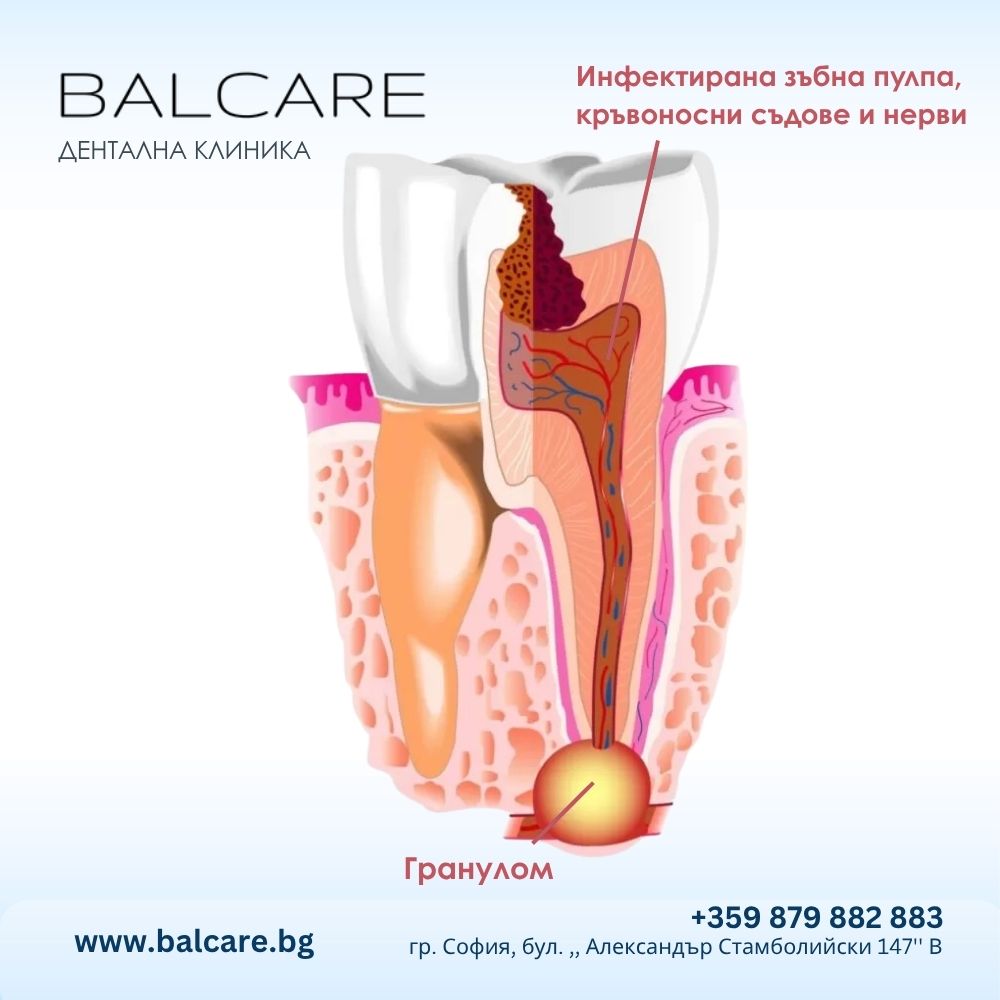
What is a dental granuloma?
Dental granuloma is a chronic inflammatory formation localized in the area of the tip of the tooth root (apex). This condition usually occurs without pronounced symptoms, which makes it difficult to detect without diagnostic imaging. The main cause of a granuloma is untreated deep caries, which leads to pulpitis (inflammation and necrosis of the dental pulp – the soft tissue containing nerves and blood vessels within the tooth).
When caries reaches the interior of the tooth and is not treated in a timely manner, bacteria penetrate the pulp and cause an inflammatory process known as pulpitis.
Over time, the inflammation progresses to nerve necrosis, leading to its complete degeneration and death. The infection does not stop there – bacteria begin to multiply in the root canals and reach the tip of the root, entering the alveolar bone – the bone that supports the teeth.
The body reacts to this invasion through an immune response, striving to limit the infection by forming an inflammatory capsule around the affected area – this is precisely what a dental granuloma is. It is a signal of a deep and often chronic infection, which must be managed through endodontic treatment under a microscope for guaranteed precision.
Types of dental granulomas
- Simple inflammatory granuloma
- Epithelial granuloma
- Cystic granuloma (radicular cyst)
This is the most common form. It forms as a result of a bacterial infection of the dental nerve, during which the immune system sends protective cells to the affected area.
These cells attempt to control the infection, creating an inflammatory reaction around the tip of the root. If left untreated, this granuloma can grow to the size of a cherry pit, leading to the destruction of the surrounding bone tissue.
This type is formed from epithelial cells that agglomerate around the zone of infection. It usually forms as a protective barrier but can progress if not treated promptly.
A cystic granuloma is a progressed form of the epithelial granuloma, in which a fluid-filled cavity forms. This cyst can reach significant sizes and cause destruction of the jawbone, leading to serious complications, including displacement or loss of teeth.
Causes, risk factors, and symptoms of granuloma
- Reasons
- Risk factors
- Symptoms
- Diagnosis
Causes of dental granuloma include:
- the presence of untreated caries, which contributes to bacterial contamination of the periodontal space (the root tip of the tooth);
incomplete or compromised dental work that allows bacteria to penetrate the dental pulp and periapical tissues; - incorrectly or incompletely performed endodontic treatment (root canal treatment) can favor the development of a chronic inflammatory formation;
- dental injuries or trauma, cracks or fractures of the tooth surface, facilitating microbial infiltration and inflammation of the periapical area.
Risk factors include:
- poor oral hygiene;
- stress;
- pressure on the teeth;
- untreated colds.
In most cases, a dental granuloma is asymptomatic.
- tooth discoloration: the affected tooth usually becomes dark in color and painful;
- pain and discomfort experienced during eating or spontaneously at rest;
- local swelling: swelling of the gums around the tooth may be observed;
- abscess: pus may form at the affected site, potentially discharging into the oral cavity and causing an unpleasant taste.
If the patient ignores the symptoms, the granuloma will increase in size and may appear as a bump next to the affected tooth. This formation often appears in a tooth known to have untreated, large-scale caries that has spread deep within.
In most cases, a dental granuloma is asymptomatic.
- tooth discoloration: the affected tooth usually becomes dark in color and painful;
- pain and discomfort experienced during eating or spontaneously at rest;
- local swelling: swelling of the gums around the tooth may be observed;
- abscess: pus may form at the affected site, potentially discharging into the oral cavity and causing an unpleasant taste.
If the patient ignores the symptoms, the granuloma will increase in size and may appear as a bump next to the affected tooth. This formation often appears in a tooth known to have untreated, large-scale caries that has spread deep within.

Treatment of granuloma
A granuloma can remain asymptomatic for many years and may flare up due to an increased bacterial load. In such cases, antibiotic therapy combined with root canal treatment of the affected tooth is indicated. If the visible part of the tooth, called the crown, is sufficiently intact to be reconstructed, treatment includes:
✅ Root canal treatment: cleaning the root canal followed by sealing with root canal filling material. A favorable result is expected about 6 months after treatment to allow the bone to recover. Some forms of chronic inflammatory lesions have a longer healing period, even up to two or three years.
The dental crown is the final stage of endodontic treatment and consists of restoring the walls destroyed by caries.
✅Tooth extraction: necessary in cases where endodontic treatment is unsuccessful and the dental granuloma remains at the same stage or, conversely, progresses.
✅Apical resection is a procedure that combines surgery with endodontics: the granuloma is surgically removed along with the affected root tip.
✅Other therapeutic methods: the laser helps reduce bacteria and sterilize the root canal. Combined with the process of mechanical cleaning and disinfection of the root canal, the laser has a high long-term success rate.
Early treatment of a dental granuloma is essential for several reasons: timely treatment prevents complications such as abscesses and cysts, relieves pain and discomfort, and preserves the affected tooth, avoiding the need for invasive procedures like root canals. It also prevents the infection from spreading to adjacent areas and into the bloodstream.
Prompt resolution of the granuloma ensures a good prognosis and lower costs compared to treating advanced infections. Delayed or postponed treatment can lead to periapical cysts and jaw defects.

Cost of granuloma treatment at BalCare Dental Clinic
The cost of granuloma treatment depends on the complexity of the case and the time required for treatment.
The primary goal in treating a dental granuloma is the effective elimination of the infection that triggers the inflammatory process in the area around the tip of the tooth root. Through precise intervention, the aim is to prevent the spread of bacteria to adjacent tissues in the oral cavity, including the gums, jawbone, and even neighboring teeth.
Patients who choose BalCare Dental Clinic for dental granuloma treatment receive not only relief from pain and the removal of discomfort but also a long-term solution that restores the aesthetics and function of the affected tooth. Thanks to modern methods such as endodontic treatment under a microscope, the clinic achieves a high success rate and a minimally invasive approach in handling even the most complex cases of granuloma.
See all prices and packages on the Prices page
At Bal Care Dental Clinic in Sofia, we offer deferred payment for root canal treatment, which facilitates access to care.
Why choose BalCare Dental Clinic for granuloma treatment?
Whether you are a teenager, a working professional, or a retired senior, your smile and dental health can be improved with quality granuloma treatment.
At BalCare Dental Clinic, we always strive to provide peace of mind to our patients. Trust your smile to the professionals.
Interest-free deferred payment for granuloma treatment.
We can provide financing for your treatment thanks to our partnership with reputable national financing organizations under favorable terms. The benefit for you is that you can start your treatment immediately.
Frequently Asked Questions About Granuloma Treatment
- What complications can arise from an untreated granuloma?
- When is apical surgery necessary?
- Is the treatment for a dental granuloma painful?
- Can the granuloma go away on its own?
- How long does the treatment take?
- How can I tell if I have a granuloma?
- Prevention
Apical granuloma can grow to visible sizes and turn into a cyst – a benign lesion that often requires surgical removal. Often, the excision of the cyst is followed by the removal of the affected root tip (apicoectomy).
Recurrence often leads to the development of an abscess with severe pain and swelling. This complication is avoided through treatment of the lesion and early diagnosis.
In some cases (especially with large granulomas or perforations), standard treatment is not enough. In such instances, apical resection is applied – the surgical removal of the root tip and the granuloma under microscopic control.
No. Thanks to modern anesthetics and microscopic equipment, the treatment is practically painless.
No. A granuloma is a chronic inflammation that does not resolve without intervention. Delay can lead to cysts and abscesses.
Usually between 1 and 2 visits, depending on the complexity.
Very often, the only way to detect it is with an X-ray, especially if there is no pain.
Prevention against the formation of granulomas is closely linked to the prevention of caries. Therefore, regular preventive check-ups, including X-rays, are the key to preventing granulomas.
Examinations should be thorough and account for asymptomatic caries and previous prosthetic work, which sometimes hide tissue devitalization – often a favorable environment for asymptomatic apical granulomas.
Applying fluoride-containing solutions to the tooth can prevent caries, while fissures, dental fillings, and root canals protect the tooth surface and prevent caries, and thus dental granulomas.
Periapical granulomas can lead to tooth loss – a situation that can easily be avoided with vigilant oral health.
