BalCare — чат


Dental Blog

Read our dental blog to learn more about your oral health, get tips on dental care, and discover helpful articles about your smile. We take pride in creating a friendly, positive dental environment where you can always feel comfortable under our care and guidance.

miofascialna-bolka-glavata-dentalna-klinika-balcare.webp
27/Apr/2026

Myofascial Head Pain: The Hidden Cause of Tension, Headaches and Disturbed Sleep – and How Red Light Therapy May Help

Myofascial pain in the head and facial region is a functional disorder that often remains undiagnosed, despite having a significant impact on patients’ quality of life. It can present with a wide range of symptoms – from diffuse facial pain and headaches to a feeling of tension, discomfort while chewing, and disturbed sleep.

In the clinical practice of BalCare Dental Clinic, these complaints are assessed in the context of myofascial pain syndrome, in which the masticatory muscles, fascial structures, and neural regulation function under conditions of chronic tension.

Not every type of head pain has the same origin, even though different conditions may feel very similar to the patient. In clinical practice, it is essential to distinguish between muscular (myofascial) pain and joint-related pain, because this determines both the underlying cause and the appropriate treatment approach.

In myofascial pain, the primary problem lies within the masticatory muscles. The muscles remain in a state of chronic tension or spasm, often as a result of teeth clenching, bruxism, or prolonged stress-related loading.

This type of pain is usually not clearly localised. It may be experienced as deep, diffuse tension and can radiate towards the temple, ear, or teeth. It is often more pronounced in the morning, when the patient wakes with a feeling of stiffness or muscular fatigue.

By contrast, joint disorders affect the intra-articular structures of the temporomandibular joint (TMJ).

In these cases, the pain tends to be more clearly localised in front of the ear and is usually triggered or intensified by movement, such as wide mouth opening or chewing.

It may also be accompanied by clicking, popping, or restricted movement, suggesting a mechanical problem within the joint itself.

This distinction has direct practical importance.

When the source of pain is muscular, the therapeutic focus is on reducing muscle tone and restoring normal function. It is in these cases that red light therapy (photobiomodulation) may be particularly beneficial, as it can support muscle relaxation, improve local circulation, and help reduce inflammatory processes.

In joint-related disorders, however, a more comprehensive approach may be required, focusing on the structures within the joint itself.

For this reason, accurate diagnosis is essential. It allows the clinician to determine not only where the pain originates, but also which therapeutic approach may be most appropriate for the individual patient.

Pathophysiology of Myofascial Pain

Myofascial pain develops as a result of prolonged muscular overload, which can lead to the formation of trigger points – localised areas of increased muscle tone and altered sensitivity.

A characteristic feature of these points is their ability to cause referred pain, meaning that pain is perceived in an area different from the actual source.

This can create diagnostic difficulties and may lead patients to consult different medical specialists without the underlying cause being identified.

Clinical Importance of the Masseter and Temporalis Muscles

The muscles most commonly involved in temporomandibular myofascial pain are the masseter and temporalis muscles, because of their central role in chewing and their tendency to become overloaded in patients with bruxism.

When trigger points are present in the masseter muscle, referred pain may be felt in the lower jaw, posterior teeth, and the area around the ear.

Patients often report increased tooth sensitivity without any objective dental findings, as well as discomfort during chewing and morning jaw stiffness.

The temporalis muscle, on the other hand, is associated with pain localised in the temple and forehead.

These symptoms are frequently interpreted as tension-type headaches without recognition of their possible muscular origin.

Trigger Points in the Masseter and Temporalis – A Common Cause of Referred Facial Pain

In clinical practice, the masseter and temporalis muscles are among the most common sources of myofascial pain in the head and facial region.

They are the primary muscles involved in closing the mouth and play an important role in clenching and bruxism.

When chronically overloaded, these muscles may develop trigger points – localised areas of increased muscle tone that can cause referred pain in different areas of the face and head.

This means that the actual source of the pain and the area where the pain is felt often do not coincide.

This mechanism helps explain why patients may report symptoms that cannot be linked to a specific structural pathology.

Masseter Muscle – Referred Pain to the Teeth, Jaw and Ear

The masseter is one of the principal muscles of mastication and is exposed to significant functional loading, particularly in patients with bruxism.

When active trigger points are present in the masseter, the following symptoms may occur:

  • pain in the lower jaw;
  • increased sensitivity in the posterior teeth without a dental cause;
  • discomfort while chewing;
  • tension and stiffness in the jaw, particularly in the morning;
  • referred pain towards the ear.

These symptoms often require differential diagnosis from odontogenic and ear, nose and throat conditions.

Temporalis Muscle – Referred Pain to the Temple and Forehead

The temporalis muscle plays an active role in stabilising the lower jaw and is particularly sensitive to chronic tension.

Trigger points in this muscle may cause:

  • pain in the temple region;
  • pain spreading towards the forehead;
  • a sensation of diffuse tension in the upper part of the face;
  • headache characterised by prolonged pressure.

This type of pain is often interpreted as a tension-type headache without consideration of its muscular origin.

Symptom Location of Pain Likely Muscular Source Clinical Characteristics
Pain in the posterior teeth without objective findings Lower jaw Masseter Intensifies during clenching or chewing
Morning stiffness and tension Jaw, cheeks Masseter Associated with nocturnal bruxism
Pain around the ear Periauricular area Masseter / pterygoid muscles Without evidence of ear pathology
Headache with diffuse pressure Temple, forehead Temporalis muscle Persistent, often bilateral
Pain during wide mouth opening Temporomandibular joint region Lateral pterygoid Intensifies with movement
Combined neck and facial pain Back of the head, jaw SCM / trapezius Associated with posture and muscular imbalance

The Relationship Between Myofascial Pain and Sleep

Chronic muscle tension may directly affect sleep quality.

Increased muscle tone and persistent pain can disturb normal sleep architecture, including through frequent micro-awakenings and a reduction in the deeper stages of sleep.

Patients often report:

  • non-restorative sleep;
  • morning fatigue;
  • headache on waking;
  • a feeling of tension in the jaw.

Scientific analyses have reported statistically significant reductions in sleep quality among patients with temporomandibular myofascial pain.

The Role of Bruxism and Stress

Myofascial pain is often associated with bruxism – involuntary clenching or grinding of the teeth, frequently occurring during sleep.

This process increases muscular loading and can contribute to the persistence of chronic pain.

Stress also plays an important role as an aggravating factor because it can increase muscle tone and activate neuromuscular mechanisms that make relaxation more difficult, even at rest.

Red Light Therapy (Photobiomodulation)

Photobiomodulation is a non-invasive therapeutic method that uses light of a specific wavelength to influence cellular processes.

Its proposed mechanisms of action include:

  • stimulation of mitochondrial activity;
  • increased ATP production;
  • improvement of local microcirculation;
  • reduction of inflammatory mediators.

As a result, pain may be reduced and muscular function may improve.

Clinical Approach at BalCare Dental Clinic

At BalCare Dental Clinic, red light therapy is used as part of a structured gnathological approach that includes functional diagnostics, assessment of muscle balance, and an individualised treatment plan.

In acute conditions, therapy is directed towards reducing pain and muscle spasm.

In chronic cases, the aim is to restore muscular function and stabilise the condition.

It is important to emphasise that photobiomodulation is not used as an isolated procedure, but as part of a comprehensive treatment approach that may include other therapeutic methods depending on the individual case.

Additional information about red light therapy:
https://balcare.bg/red-light-therapy/

Myofascial pain in the head and facial region is a complex condition that affects not only the local musculature but also sleep quality, neural regulation, and the patient’s everyday comfort.

Recognition of trigger points and accurate differential diagnosis are essential for effective treatment.

Modern therapeutic methods, including photobiomodulation, offer the possibility of targeting key pain mechanisms when applied according to an appropriate protocol and as part of a comprehensive treatment strategy.

Bibliography

https://pmc.ncbi.nlm.nih.gov/articles/PMC8291152/
Systematic review and meta-analysis on the effectiveness of photobiomodulation for myofascial pain associated with temporomandibular disorders.

https://pubmed.ncbi.nlm.nih.gov/29697718/
Systematic review of low-level light therapy for temporomandibular myofascial pain and its relationship with symptoms.

https://link.springer.com/article/10.1186/s12903-023-02784-8
Randomised clinical trial comparing the effects of LED and laser therapy in patients with temporomandibular disorders.

https://pmc.ncbi.nlm.nih.gov/articles/PMC7356229/
Review article on the cellular mechanisms of photobiomodulation.


temporomandibularna-stava-syn-denatalna-klinika-balcare.webp
22/Apr/2026

How Does the Temporomandibular Joint Affect Sleep?

Problems involving the temporomandibular joint (TMJ) are often viewed simply as local discomfort in the jaw area. In clinical practice, however, something much more significant is increasingly being observed – a direct relationship between temporomandibular disorders, chronic pain, and sleep disturbances.

Patients rarely associate morning fatigue, frequent awakenings, or nighttime teeth clenching with a problem involving the jaw joint, which may delay seeking appropriate treatment.

The temporomandibular joint is a small but extremely important structure that connects the lower jaw to the skull. It is involved every time we open the mouth, chew, speak, yawn, or swallow.

When this joint or the muscles surrounding it do not function properly, the problem rarely remains limited to “jaw pain”. Many patients begin to experience morning headaches, facial tension, pain around the ear, tooth sensitivity, stiffness when opening the mouth, and disturbed sleep.

At BalCare Dental Clinic, temporomandibular dysfunction is approached as a functional problem that may affect not only the bite and the masticatory muscles, but also sleep quality.

This is particularly important for patients who wake feeling tired, clench their teeth during the night, or feel that they are “unable to relax their jaw”.

Why Can a Jaw Problem Disturb Sleep?

Many patients do not associate poor sleep with their teeth or jaw.

They may look for the cause in stress, exhaustion, hormonal changes, an uncomfortable pillow, or everyday tension.

These factors may indeed contribute, but temporomandibular dysfunction introduces another important mechanism: the body may be unable to relax fully because the masticatory muscles remain active during the night.

When the masseter, temporalis, and other muscles surrounding the joint remain tense, the brain continues to receive signals of discomfort.

This may contribute to micro-awakenings that the patient does not remember the following morning but experiences indirectly as fatigue, irritability, and a lack of restorative sleep.

The Most Common Symptoms Patients Tend to Ignore

Temporomandibular dysfunction does not always begin with acute pain.

The first signs are often much more subtle:

  • morning jaw tension;
  • facial pain or fatigue;
  • headaches after waking;
  • clicking or popping when opening the mouth;
  • difficulty chewing;
  • pain around the ear without an identifiable ear-related cause;
  • teeth clenching or grinding;
  • tooth sensitivity;
  • disturbed, light, or fragmented sleep.

These symptoms are often treated separately – with painkillers, massage, a new pillow, or sleep supplements.

However, when the underlying cause involves the masticatory system, the relief is often only temporary.

Bruxism: Nighttime Clenching That Can Maintain the Problem

Bruxism is one of the important factors in patients with temporomandibular disorders and disturbed sleep.

It involves clenching or grinding of the teeth, often occurring unconsciously during sleep. This places additional load on the temporomandibular joint, masticatory muscles, and teeth.

The problem is that bruxism may create a vicious cycle.

The more a patient clenches their teeth, the more the muscles become overloaded. The greater the muscular tension, the more pronounced the pain may become. And the stronger the discomfort, the more difficult it may be for the body to reach deep, restorative sleep.

An important professional distinction should be made here: not every case of bruxism indicates severe temporomandibular dysfunction.

However, when bruxism is accompanied by pain, morning stiffness, headaches, and disturbed sleep, a gnathological assessment is recommended.

The Relationship Between TMD and Sleep Apnoea – What Patients Should Know

Sleep apnoea is a condition in which breathing repeatedly stops or becomes significantly reduced during sleep.

The most common form is obstructive sleep apnoea (OSA), in which the upper airway becomes narrowed or temporarily blocked.

This may lead to:

  • snoring;
  • frequent awakenings;
  • dry mouth in the morning;
  • daytime sleepiness;
  • headaches;
  • increased cardiovascular risk.

The relationship between temporomandibular disorders and sleep apnoea is complex and should not be oversimplified.

It would not be accurate to claim that TMD “causes” sleep apnoea in every patient.

A more precise explanation is that some patients may have overlapping factors, including:

  • the position of the lower jaw;
  • muscular tension;
  • bruxism;
  • disturbed breathing during sleep;
  • unstable or fragmented sleep.

More recent studies and review publications have reported an association between painful temporomandibular disorders and obstructive sleep apnoea, although the scientific evidence still requires careful interpretation.

A 2021 review indicated that the evidence supporting an association between TMD and sleep quality is stronger, while the relationship between TMD and sleep apnoea requires further high-quality research.

A more recent meta-analysis published in 2024 reported a significant association between TMD and obstructive sleep apnoea.

The practical message for patients is clear:

If jaw pain is accompanied by loud snoring, pauses in breathing, sudden awakenings with shortness of breath, excessive daytime sleepiness, or high blood pressure, consultation with a sleep medicine specialist or an ear, nose and throat (ENT) specialist is also necessary.

The role of the dentist and gnathologist is to assess jaw function, the bite, muscular tension, and the possible need for dental appliances.

However, sleep apnoea requires appropriate medical diagnosis.

How Does Red Light Therapy Fit Into Treatment?

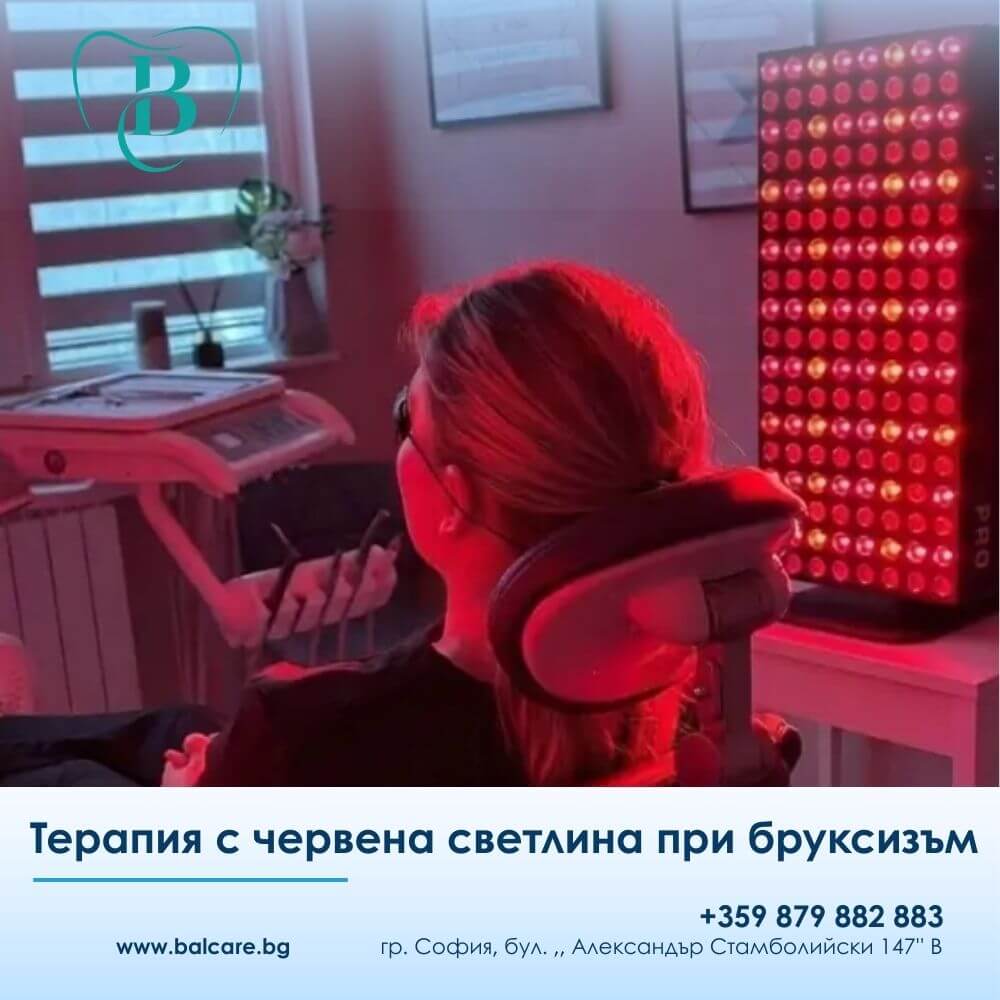
Red light therapy, also known as photobiomodulation, is a modern non-invasive treatment method that uses red and/or near-infrared light with specific therapeutic parameters.

Its purpose is not simply to “warm” the area or provide temporary relaxation.

Instead, the therapy is intended to influence cellular processes, local circulation, inflammation, and tissue recovery.

In patients with temporomandibular dysfunction, therapy may be directed towards the joint area and the masticatory muscles when indicated after examination.

At BalCare Dental Clinic, photobiomodulation is not applied randomly or according to the principle that “more is better”.

Treatment follows specific protocols, with defined treatment areas, duration, and sequence of procedures according to the patient’s individual condition.

This distinction is important because photobiomodulation is a dose-dependent therapy.

Its effects depend on appropriate assessment, the correct area of application, and the correct treatment protocol.

A 2023 systematic review on photobiomodulation for temporomandibular disorders reported promising effects in reducing pain and improving maximum mouth opening.

How Can Reducing Pain Improve Sleep?

Red light therapy is not a sleeping medication.

It does not act as a drug for insomnia and should not be presented in this way.

Its potential value for patients with temporomandibular disorders lies in helping to reduce some of the factors that may interfere with sleep:

  • pain;
  • inflammation;
  • muscular tension.

When the masticatory muscles become more relaxed, it may be easier for the patient to reach a state of rest.

When pain is reduced, there may be fewer pain-related micro-awakenings.

When the joint is less irritated, morning stiffness and discomfort may also decrease.

This is the practical relationship between TMD, red light therapy, and sleep:

The treatment does not directly target “insomnia”; instead, it may help address one of the potential sources of nighttime discomfort.

The Approach at BalCare Dental Clinic

At BalCare Dental Clinic, red light therapy is considered part of a comprehensive treatment strategy rather than a universal stand-alone solution.

For patients experiencing jaw pain, bruxism, and disturbed sleep, the first step is to assess the function of the masticatory system.

The treatment plan may include:

  • gnathological assessment;
  • bite evaluation;
  • examination of the masticatory muscles;
  • assessment of the temporomandibular joint;
  • red light therapy according to an individual protocol;
  • a custom-made occlusal splint for bruxism when indicated;
  • guidance for reducing muscular overload;
  • follow-up assessment of treatment results.

What Is Important Not to Overlook?

One of the most important aspects often missing from discussions of this subject is the distinction between a symptom and its underlying cause.

Jaw pain may result from:

  • muscular overload;
  • abnormal functional loading;
  • bruxism;
  • a joint disorder;
  • stress;
  • sleep disturbances;
  • or a combination of several factors.

For this reason, treatment must be individualised.

In one patient, the primary problem may be nighttime clenching.

In another, it may be inflammation and pain within the joint.

A third patient may have sleep apnoea, in which case the dental approach needs to be coordinated with appropriate medical diagnosis.

For another patient, an unstable bite and chronic overloading of specific muscles may be the main contributing factors.

The professional approach is not to promise a quick universal solution, but to identify the precise mechanism behind the patient’s symptoms.

When Should You Seek a Consultation?

A professional assessment is recommended if you experience:

  • jaw pain in the morning;
  • teeth clenching or grinding;
  • frequent headaches;
  • clicking or popping in the joint;
  • facial tension;
  • pain around the ear;
  • fragmented sleep;
  • fatigue despite apparently sufficient hours of sleep;
  • snoring or suspected sleep apnoea.

When such symptoms are present, assessment should not be unnecessarily delayed.

The earlier the underlying cause is identified, the easier it may be to interrupt the cycle of:

pain – muscle tension – bruxism – disturbed sleep.

The temporomandibular joint may have a greater relationship with sleep quality than many patients realise.

When the jaw remains tense, the muscles are overloaded, and the joint is irritated, the resulting discomfort may make restorative sleep more difficult.

Scientific evidence supports an association between temporomandibular disorders and impaired sleep quality.

Photobiomodulation, or red light therapy, is a modern therapeutic method that may support the reduction of pain and muscular tension when it is appropriately indicated and applied according to a defined protocol.

At BalCare Dental Clinic, the approach is based on comprehensive assessment of the masticatory system, an individual treatment plan, and monitoring of the results.

The goal is not simply temporary symptom relief, but the restoration of function, comfort, and quality of life.


kakvo-predstavlyava-red-light-therapy-dentalna-klinika-balcare.webp
06/Apr/2026

What Is Red Light Therapy?

How to Prepare for Your First Session at BalCare Dental Clinic

What to expect: protocols, duration, sensations, and realistic results

At BalCare Dental Clinic, Red Light Therapy is used as a modern, gentle, and effective method for relieving jaw pain, bruxism, and muscular tension.

Many patients want to know what happens during the first session and what they can realistically expect from the therapy.

This article provides clear and practical information to help you prepare with confidence and get the maximum benefit from the treatment.

Stimulating Cellular Activity

Red Light Therapy, also known as photobiomodulation, uses specific wavelengths of red and near-infrared light that penetrate the tissues and stimulate cellular activity.

This may contribute to:

  • reducing inflammation;
  • supporting tissue recovery;
  • relaxing the muscles;
  • reducing pain.

In dentistry, the method is used for conditions such as:

  • jaw pain;
  • temporomandibular dysfunction;
  • bruxism and teeth clenching;
  • muscular tension.

If you experience similar symptoms, you can also read our detailed article about jaw pain.

How to Prepare for Your First Session

Preparation is simple, but it can be important for the effectiveness of the therapy.

It is recommended that:

✔️ no creams or make-up are applied to the facial treatment area;
✔️ you inform the clinician about any medications you are taking and any relevant medical conditions;
✔️ you mention if you use an occlusal splint for bruxism.

No fasting or special preparation is required.

It is best to arrive as relaxed as possible, as stress can influence muscle tone.

What Happens During the First Appointment – Step by Step

The first appointment begins with a clinical assessment of your condition.

The clinician evaluates:

✔️ muscular tension;
✔️ painful or sensitive points;
✔️ range of motion;
✔️ temporomandibular joint function.

An individual treatment protocol is then prepared according to the specific problem.

The patient is positioned comfortably, protective glasses are provided, and the light device is activated.

How Long Does the First Session Take?

The procedure usually lasts approximately 10 to 12 minutes per side.

For milder conditions, treatment may be shorter, while chronic conditions may require longer treatment and application to several areas.

What Will You Feel During the Therapy?

The therapy is completely painless.

The most common sensations include:

✔️ gentle warmth;
✔️ relaxation of the muscles;
✔️ an overall feeling of relaxation.

Some patients notice a reduction in tension even during the first treatment session.

What Results Can You Expect After the First Session?

After the first procedure, some patients may experience:

✔️ reduced pain;
✔️ less tension in the jaw;
✔️ greater comfort while chewing.

With consecutive sessions, the effect may gradually build and stabilise.

If you experience nighttime teeth clenching, you can also read: Red Light Therapy for Bruxism.

How Many Sessions Are Needed?

✔️ 12 sessions for muscular problems
✔️ 24 sessions for joint-related problems

Is Red Light Therapy Safe?

Yes, the therapy is safe and gentle when applied correctly.

✔️ No medication is used.
✔️ The tissues are not damaged.
✔️ When properly applied, the treatment is designed to minimise unwanted effects.

Are There Any Contraindications?

In certain situations, prior consultation is necessary, including:

✔️ photosensitivity;
✔️ use of certain medications;
✔️ oncological conditions;
✔️ pregnancy.

When Is Red Light Therapy Not Enough on Its Own?

Some conditions require a combined treatment approach.

Red Light Therapy may be combined with:

  • an occlusal splint for bruxism;
  • orthodontic treatment;
  • bite correction.

Can It Be Combined with Other Treatments?

Yes.

Red Light Therapy can be combined with different dental and restorative treatments and may support the recovery process.

What Should You NOT Do After the Procedure?

There are no specific restrictions required after the treatment.

Comparison with Other Methods

  • Medication – temporary symptom relief;
  • occlusal splints – protection of the teeth;
  • Red Light Therapy – targets the underlying muscular and inflammatory processes.

A combined approach may provide the best results.

Frequently Asked Questions

How Quickly Does Red Light Therapy Work?

Some patients may experience relief after the first session, although the best results are usually achieved after a series of treatments.

Is the Procedure Painful?

No. The therapy is completely painless.

Is It Suitable for Bruxism?

Yes. It can support muscle relaxation and help reduce muscular tension.

How Long Does the Effect Last?

With an appropriate protocol and maintenance, the effects may be long-lasting.

At BalCare Dental Clinic, Red Light Therapy is applied using an individual approach for every patient.

A personalised protocol is prepared according to the patient’s condition, symptoms, and the functional load on the masticatory system.

A significant reduction in pain, muscular tension, and discomfort may be observed in patients with bruxism and temporomandibular disorders when therapy is performed consistently and combined with appropriate habits and additional treatment when necessary.

Red Light Therapy is a modern therapeutic method that may provide noticeable relief from the first sessions.

The first session represents the beginning of a recovery process aimed at improving jaw function and quality of life.

If you experience pain, muscular tension, or bruxism, Red Light Therapy may be an important step towards a longer-term solution.


red-light-therapy-bruxism-dentalna-klinika-balcare.jpg
18/Mar/2026

Red Light Therapy for Bruxism – A Modern Approach to Relieving Pain and Muscle Tension

Teeth grinding is not simply a habit. It is a condition that often goes unnoticed until the first symptoms appear – jaw tension, pain, or morning headaches.

In clinical practice, we often see patients who ignore these signs for a long time until the discomfort becomes persistent.

Bruxism is, in fact, a complex condition. The masticatory muscles remain under increased tension, the tissues are repeatedly overloaded, local circulation may be affected, and inflammatory processes may gradually develop.

These mechanisms can contribute to symptoms such as jaw tightness, pain when opening the mouth, tooth sensitivity, and frequent headaches.

If the condition is not addressed in time, the symptoms may become more pronounced and develop into a chronic problem.

How Red Light Therapy Affects Pain

Red Light Therapy, also known as photobiomodulation, is a modern non-invasive method that works at the cellular level.

The light penetrates the tissues without damaging them and stimulates cellular processes, including mitochondrial activity – an important part of cellular energy production.

As a result, cellular energy production may increase, supporting natural recovery processes.

This may contribute to:

  • reducing inflammation;
  • relieving pain;
  • improving local circulation;
  • supporting tissue recovery.

This mechanism is one of the reasons photobiomodulation is used in different areas of dental medicine, including the management of oral pain, inflammatory conditions, and muscular tension.

What Results Can Patients Expect?

Clinical observations and current research suggest that Red Light Therapy may help reduce pain in the jaw and oral region, particularly in patients with temporomandibular disorders that may coexist with bruxism.

In addition to pain relief, the therapy may also influence inflammatory processes within the tissues.

This can be particularly relevant in conditions involving irritated soft tissues, gingival inflammation, or minor tissue trauma associated with prolonged clenching.

Another important potential benefit is support for tissue recovery.

Stimulation of cellular activity may assist soft-tissue regeneration and reduce discomfort following certain dental procedures.

For this reason, the method can be valuable not only in patients with bruxism but also as part of a broader therapeutic approach.

The effect on the patient’s general well-being should not be underestimated either.

Oral and jaw pain can interfere with sleep, eating, and everyday comfort. When the pain is brought under better control, both physical comfort and overall well-being may improve.

How Does It Help Specifically with Bruxism?

In patients with bruxism, Red Light Therapy may have a beneficial effect on the muscles and temporomandibular joint region.

It may help:

  • relax the masticatory muscles;
  • reduce morning stiffness;
  • relieve tension in the temple area.

At the same time, improved local circulation may support recovery after prolonged muscular overload.

It is important to understand that Red Light Therapy does not eliminate the behaviour of clenching or grinding itself.

Its role is primarily to reduce associated symptoms and improve day-to-day comfort.

How Is the Therapy Performed?

The procedure is completely non-invasive and comfortable for the patient.

The light source is directed towards the jaw area or other affected regions, with one session usually lasting approximately 10 to 20 minutes.

The treatment is painless.

Patients most commonly describe a mild sensation of warmth and gradual relaxation of the muscles, which can make the procedure feel calming and comfortable.

How Long Does It Take to Notice an Effect?

The effects of therapy are generally cumulative rather than immediate.

Initial improvement may be noticed after several sessions, while a more noticeable effect is often achieved after a series of approximately 6 to 10 treatments.

In chronic conditions, maintenance sessions may sometimes be recommended to help preserve the achieved result.

An Important Point Patients Should Understand

Red Light Therapy should not be considered a stand-alone solution.

The best results are generally achieved when it forms part of a comprehensive treatment plan.

In practice, this may mean combining the therapy with:

  • a custom-made night-time occlusal splint to protect the teeth;
  • stress management;
  • bite correction when clinically indicated.

Bruxism and the associated pain in the mouth and jaw should not simply be accepted as normal.

They are signs of functional overload that deserve proper assessment and a professional treatment approach.

Red Light Therapy is a modern, non-invasive therapeutic method that supports the body’s natural recovery processes.

It may help reduce pain, relax overloaded muscles, and improve quality of life – without medication and without invasive procedures.

When appropriately indicated and combined with other treatment methods, it can become an important component in the successful management of symptoms associated with bruxism.


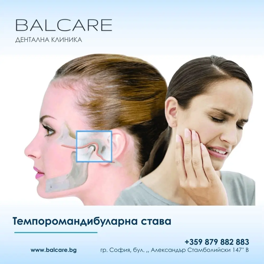
temporomandibularna-stava-dentalna-klinika-balcare.webp
15/Feb/2026

What Is the Temporomandibular Joint?

The temporomandibular joint (TMJ) is a paired joint that connects the lower jaw to the skull. It is located just in front of the ears on both sides of the head and is one of the most complex joints in the human body.

Every time we speak, chew, yawn, or swallow, we use the temporomandibular joint.

The TMJ functions partly like a hinge, allowing the jaw to move up and down, forward and backward, and from side to side. This flexibility enables us to speak, chew, and express emotions through facial movements.

Anatomy of the Temporomandibular Joint

The temporomandibular joint consists of several key components:

  • Joint capsule – surrounds the joint and helps maintain its stability;
  • Articular disc – a cartilaginous structure that acts as a cushion between the bony surfaces;
  • Muscles – provide and control jaw movement;
  • Ligaments – support the joint and help limit excessive movement.

When all of these components work together harmoniously, the joint functions smoothly and without pain.

The Most Common Symptoms and Complaints

Problems involving the temporomandibular joint, known as temporomandibular disorders (TMD), can cause a wide range of symptoms.

Pain and Discomfort

  • dull or sharp jaw pain;
  • pain around the ear, which may sometimes be mistaken for an ear infection;
  • pain in the temples and cheeks;
  • headaches, particularly in the temple region;
  • pain in the neck and shoulders.

Joint Sounds and Movement Problems

  • clicking or popping when opening or closing the mouth;
  • grating or grinding sounds within the joint;
  • a feeling that the jaw “catches” or “locks”;
  • restricted mouth opening;
  • deviation of the jaw during opening.

Other Symptoms

  • difficulty chewing or discomfort while eating;
  • dizziness or vertigo;
  • ringing in the ears (tinnitus);
  • a sensation of blocked or full ears;
  • changes in the way the upper and lower teeth come together (occlusion).

What Causes Temporomandibular Disorders?

Temporomandibular disorders may have multiple causes, and several factors are often present at the same time.

Physical Factors

  • Bruxism – grinding or clenching the teeth, particularly during sleep;
  • Trauma – injury or impact affecting the jaw or head;
  • Arthritis – inflammatory disease involving the joint;
  • abnormal occlusion or tooth contacts;
  • degenerative or structural changes involving the articular disc;
  • poor posture, particularly involving the head and neck.

Habits and Lifestyle Factors

  • chewing gum for prolonged periods;
  • biting hard objects such as pencils or nails;
  • chewing predominantly on one side;
  • resting the jaw on the hand;
  • poor workplace ergonomics.

Emotional Factors

  • Stress – one of the important contributing factors;
  • anxiety;
  • depression;
  • tension that contributes to jaw clenching.

How Are Temporomandibular Disorders Diagnosed?

Diagnosis of temporomandibular disorders generally involves several steps.

Clinical Examination

The dental professional will:

  • take a detailed history of the patient’s symptoms;
  • examine jaw movement;
  • palpate the muscles and joint for tender or painful areas;
  • assess how the teeth come together;
  • check for joint sounds.

Additional Diagnostic Tests

When necessary, further imaging may be recommended:

  • X-ray – for assessment of bony structures;
  • Magnetic resonance imaging (MRI) – for visualisation of soft tissues and the articular disc;
  • Computed tomography (CT) – for detailed evaluation of the bone.

Treatment Options

Treatment for temporomandibular disorders is individualised and depends on the cause and severity of the condition.

Conservative Treatment – First-Line Approach

Many cases improve with non-invasive methods.

Self-Care and Habit Modification

  • avoiding very hard foods;
  • taking smaller bites;
  • avoiding extreme jaw movements, including excessive yawning;
  • applying warm or cold compresses;
  • relaxation and stress-management techniques.

Physiotherapy

  • jaw strengthening exercises;
  • stretching exercises;
  • muscle massage;
  • LED red light therapy.

Medication

When clinically appropriate, treatment may include:

  • anti-inflammatory medication such as ibuprofen or naproxen;
  • muscle relaxants;
  • pain-relieving medication when needed;
  • in selected cases, antidepressant medication for chronic pain management.

Dental Treatment

Occlusal Splint

A custom-made occlusal splint may be used to:

  • protect the teeth from grinding and clenching;
  • reduce excessive loading on the joint;
  • support a more stable jaw position;
  • provide an individually fabricated appliance made by a dental laboratory.

Orthodontic Treatment

Orthodontic treatment may be considered when correction of the bite is necessary.

It can help improve tooth alignment and occlusal relationships when clinically indicated.

Prosthodontic Treatment

This may include:

  • replacement of missing teeth;
  • restoration of severely worn teeth.

Prevention and Care of the Temporomandibular Joint

Everyday Habits

  • Maintain good posture – keep your head in a balanced position, particularly when working at a computer;
  • Avoid unnecessary teeth clenching – the jaw should remain relaxed when you are not eating or speaking;
  • Chew on both sides – distribute the functional load as evenly as possible;
  • Limit chewing gum – avoid prolonged gum chewing;
  • Be careful when yawning – support the jaw if you need to open the mouth very widely.

Stress Management

  • practise relaxation techniques such as meditation and deep breathing;
  • get sufficient sleep;
  • maintain regular physical activity;
  • consider yoga or tai chi;
  • seek psychological support when appropriate.

Dietary Recommendations

  • avoid very hard foods such as nuts, ice, and hard sweets during painful periods;
  • cut food into smaller pieces;
  • choose softer foods during a flare-up;
  • avoid taking very large bites.

Frequently Asked Questions

Is Clicking in the Jaw Dangerous?

Clicking on its own is not necessarily dangerous.

Many people experience joint clicking without pain or other symptoms.

However, if the clicking is accompanied by pain, restricted movement, locking, or other symptoms, consultation with a specialist is recommended.

How Long Does Treatment Take?

The duration of treatment varies considerably depending on the severity of the problem and the individual response to therapy.

Mild cases may improve within several weeks, while chronic conditions may require several months or longer-term management.

Will I Need to Wear a Splint for the Rest of My Life?

Not necessarily.

Many patients use an occlusal splint temporarily while their symptoms improve and while new habits are established.

However, some patients may require longer-term use, particularly when chronic bruxism is present.

Can Temporomandibular Disorders Be Completely Treated?

Many patients achieve significant or complete relief from their symptoms with appropriate treatment.

For others, TMD may be a chronic condition that requires long-term management.

Early diagnosis and an appropriate individual treatment plan are important.

Will I Need Surgery?

Most temporomandibular disorders can be successfully managed without surgery.

Surgical treatment is generally considered only in selected severe cases when appropriate conservative treatment has not produced sufficient improvement.

Does Stress Really Affect the Temporomandibular Joint?

Yes.

Stress can be an important contributing factor.

When people are under stress, they may unconsciously clench or grind their teeth, increasing the load on the temporomandibular joint and the surrounding muscles.

For this reason, stress management can be an important part of treatment.

Can I Chew Gum?

If you have problems involving the temporomandibular joint, it is generally advisable to limit or temporarily avoid chewing gum because it increases repetitive loading of the joint.

Once the symptoms improve, moderate gum chewing may be resumed if your dental professional considers it appropriate.

When Should I Seek Professional Help?

You should consider consulting a specialist if:

  • you have persistent or frequently recurring jaw pain;
  • you cannot fully open or close your mouth;
  • significant clicking or popping is accompanied by pain;
  • your symptoms interfere with eating, speaking, sleeping, or your overall quality of life.

Is There a Relationship Between Dental Procedures and Temporomandibular Disorders?

Long or complex dental procedures may temporarily aggravate TMD symptoms because the mouth remains open for an extended period.

If you already have problems involving the temporomandibular joint, inform your dentist before treatment so that appropriate precautions can be taken.

The temporomandibular joint plays a vital role in everyday life, even though most of us rarely think about it.

Problems involving the TMJ can significantly affect quality of life, but with timely diagnosis and appropriate treatment, many patients experience substantial improvement.

If you have symptoms associated with the temporomandibular joint, do not wait for them to become more severe.

Early assessment and treatment are often the most effective way to prevent persistent or chronic problems.

For a consultation and examination, contact Dr Trajko Balabanov, who provides an individual approach to the diagnosis and treatment of conditions related to the temporomandibular joint.

Do not let jaw pain limit your daily life. Book a consultation today.


kak-deystvat-alaynerite-dentalna-klinika-balcare.webp
25/Jan/2026

How Do Clear Aligners Work?

Misaligned teeth are not only an aesthetic concern. Malocclusions can lead to functional problems, accelerated tooth wear, gum inflammation, temporomandibular joint discomfort, and difficulties maintaining proper oral hygiene.

In recent years, treatment with clear aligners has become established as an evidence-based and minimally invasive method of orthodontic correction, particularly among adult patients.

As an orthodontist with more than ten years of clinical experience, I would like to emphasise one important point from the outset: aligners are not a cosmetic accessory. They are medical devices that require comprehensive diagnostics, digital treatment planning, and continuous professional supervision.

What Are Clear Aligners?

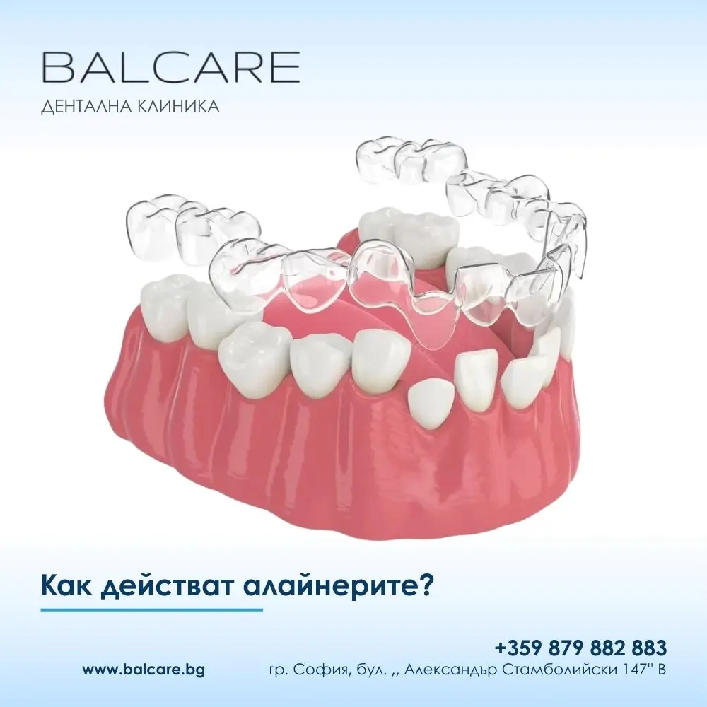
Aligners are thin, transparent, custom-made orthodontic trays that fit closely over the teeth.

They are manufactured from specialised medical-grade polymers and are worn according to a precisely prescribed schedule.

Their main function is to apply controlled, carefully calculated pressure to the teeth, gradually moving them into predetermined positions.

The word aligner itself refers to the process of alignment, which accurately describes their role in orthodontic treatment.

The Biological Principle: How Do Teeth Move?

Each tooth is anchored within the jawbone by the periodontal ligament – a living tissue that responds to mechanical forces.

When a constant, gentle, and properly controlled force is applied to a tooth, biological remodelling of the surrounding bone takes place.

Clear aligners use this natural biological mechanism.

Each aligner moves the teeth by a very small increment – usually approximately 0.2–0.3 mm.

This allows tooth movement to be controlled, predictable, and gentle on the roots and surrounding tissues.

Clear Aligner Treatment – Step by Step

1. Clinical and Functional Diagnosis

Treatment begins with a comprehensive orthodontic examination, evaluation of the bite, assessment of the gums and supporting bone, and radiographic diagnostics when necessary.

At this stage, the orthodontist determines whether the individual case is suitable for clear aligner therapy.

2. Digital 3D Treatment Planning

An intraoral scanner or highly accurate dental impressions are used to create a digital model of the teeth.

Specialised software allows every stage of tooth movement to be planned – from the initial position to the intended final result.

The patient can view a digital simulation of the treatment before the active phase begins.

3. Production of Custom Aligners

Based on the digital treatment plan, a series of sequential aligners is produced.

Each new tray has a slightly modified shape designed to guide the teeth towards the next planned position.

4. Wearing the Aligners and Follow-Up

Aligners are usually worn for 20–22 hours per day and removed only for eating and oral hygiene.

Follow-up appointments are generally scheduled every 6–8 weeks to monitor progress and make adjustments when necessary.

How Long Does Clear Aligner Treatment Take?

Treatment duration depends on the complexity of the case, the patient’s biological response, and compliance with the recommended wearing schedule.

Case Complexity Average Treatment Duration
Mild corrections 3–6 months
Moderate complexity 6–12 months
Complex cases 12–24 months

What Dental Problems Can Be Treated with Aligners?

Clear aligners can be effective for the treatment of:

  • crowded teeth;
  • gaps between teeth;
  • tilted teeth;
  • rotated teeth;
  • crossbite;
  • certain cases of open bite.

When appropriately indicated, more than 70–80% of adult patients may be successfully treated with clear aligners.

Limitations and Contraindications

Clear aligners are not a universal solution.

They may not be appropriate in cases involving:

  • severe skeletal discrepancies;
  • significant vertical bite problems;
  • active periodontal disease;
  • uncontrolled bruxism;
  • cases requiring orthognathic surgery.

An honest assessment of these limitations is an essential part of professional orthodontic practice.

Attachments and Auxiliary Components – Why Are They Needed?

In many cases, small composite elements known as attachments are bonded to the teeth.

They act as anchorage points that allow the aligners to achieve more complex movements, including:

  • rotations;
  • uprighting tilted teeth;
  • improved control of tooth roots.

In some cases, treatment may also be supported by additional orthodontic components such as:

  • temporary anchorage devices or mini-implants;
  • springs;
  • transpalatal arches.

These auxiliary elements can support more complex orthodontic movements without compromising the overall aesthetic advantages of aligner treatment.

Clear Aligners or Braces?

Criterion Clear Aligners Fixed Braces
Aesthetics Almost invisible Visible
Oral hygiene Easier to maintain More challenging
Comfort High Generally lower
Patient compliance Highly dependent on discipline Less dependent on patient compliance
Complex tooth movements May require auxiliary elements Often easier to achieve

Possible Risks and Side Effects

Even when treatment is properly planned and carried out, some temporary effects may occur:

  • temporary tooth sensitivity;
  • mild changes in speech at the beginning of treatment;
  • tension or pressure in the jaw;
  • risk of relapse if retention is not followed.

These effects are generally temporary and manageable.

What Happens If the Recommended Wearing Schedule Is Not Followed?

Failure to wear the aligners as prescribed may result in:

  • loss of control over the planned tooth movements;
  • prolonged treatment time;
  • the need for new digital planning;
  • additional treatment costs.

Patient compliance is one of the key factors determining the success of aligner treatment.

Care and Hygiene of Clear Aligners

Aligners should be cleaned with a soft brush and water.

Abrasive toothpastes are not recommended because they may scratch the material and make the trays appear cloudy.

Special aligner-cleaning tablets may be used when necessary.

Whenever the aligners are removed, they should be stored in a protective case.

Retention – The Essential Final Stage of Treatment

After the active phase of orthodontic treatment, a retainer must be used.

The retainer stabilises the achieved result and helps prevent the teeth from moving back towards their original positions.

Retention is an essential and inseparable part of orthodontic treatment.

Clear aligners are a high-tech, effective, and minimally invasive orthodontic solution when they are properly indicated, professionally planned, and worn according to instructions.

They can provide predictable results, excellent aesthetics, and a high level of comfort, but successful treatment requires expert supervision and patient cooperation.

A straight smile is not only about appearance – it is an important part of overall dental and functional health.


alaineri-vyzrastni-dentalna-klinika-balcare.webp
16/Jan/2026

Clear Aligners for Adults – A Modern Orthodontic Solution for a Discreet and Healthy Smile

What are clear aligners for adults, and why is there so much interest in them?

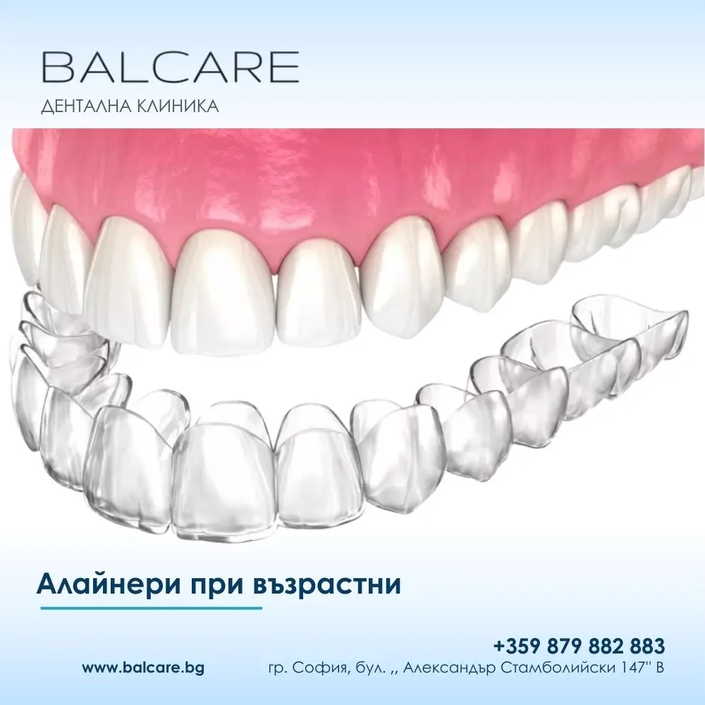
Clear aligners for adults are a modern orthodontic treatment method that combines aesthetics, comfort, and clinically established effectiveness. They are transparent, removable trays created through digital planning and 3D technology, gradually moving the teeth into the desired position.

The pressure applied to the teeth is calculated in advance using specialised digital orthodontic software and changes with each successive aligner.

Each aligner moves the teeth by approximately 0.2–0.3 mm, allowing for gradual and well-controlled movement. This progressive approach is one of the reasons treatment is generally well tolerated and associated with minimal discomfort.

More and more people over the age of 25, 35, and even 50 are choosing aligners because they want to improve their smile without visible braces and without compromising their professional or social life.

Modern orthodontics is no longer limited by age. What matters most is the biological condition of the tissues and appropriate treatment planning.

In this article, we answer the questions adult patients most commonly ask: Is treatment possible? How do aligners work? How long does treatment take? What determines the cost? What are the advantages and limitations? And when may aligners be a better choice than braces?

The aim is to provide clear, useful, and structured information that supports an informed treatment decision.

Is Orthodontic Treatment with Aligners Possible for Adults?

Yes. Orthodontic treatment with clear aligners is entirely possible in adults and is well established in clinical practice, provided that the gums and supporting bone are healthy.

Unlike in children, tooth movement in adults may sometimes occur more slowly, but it remains predictable when the correct biomechanical principles are applied.

Current research shows that bone remodelling around the teeth continues throughout life.

This means that age itself is not a contraindication. The key factors are periodontal health and compliance with the prescribed wearing schedule.

https://www.youtube.com/watch?v=xsP7N1H5xQs

Why Are Clear Aligners Particularly Suitable for Adults?

Clear aligners respond directly to the needs of adult patients because they combine functionality with discretion.

They are almost invisible, which is particularly important for people whose professional or social life involves regular face-to-face communication.

They are also removable, making eating and maintaining excellent oral hygiene easier.

Clinical observations indicate that adult patients treated with aligners may have a lower risk of tooth decay and gingival inflammation compared with patients wearing fixed orthodontic appliances.

What Problems Can Be Corrected with Clear Aligners in Adults?

Aligners can be effective for a wide range of dental and orthodontic problems, particularly mild to moderate cases.

They are commonly used for:

  • crowded teeth;
  • diastemas and other spaces between teeth;
  • rotated teeth;
  • tilted teeth;
  • tooth migration following tooth loss.

In more complex cases, including pronounced skeletal discrepancies, aligners may form part of a combined treatment approach involving other orthodontic techniques.

The decision is always based on individual diagnosis.

Advantages of Clear Aligners for Adults

Clear aligners provide excellent aesthetics throughout treatment, with no visible metal components and less irritation of the soft tissues.

They allow patients to eat normally, maintain oral hygiene more easily, and in many cases require fewer orthodontic appointments.

Another important advantage is the predictability provided by digital treatment planning.

Digital technology allows precise control over each stage of tooth movement, which can be particularly valuable for adult patients with busy schedules.

Disadvantages and Limitations of Clear Aligner Treatment

The main limitation of aligner treatment is the need for a high level of patient discipline.

If the trays are not worn for the recommended number of hours, treatment may take longer or the planned result may be compromised.

In severe dental or skeletal discrepancies, aligners may not be sufficient as a stand-alone treatment and a combined approach may be required.

This is not a weakness of the method, but part of sound orthodontic decision-making.

Clear Aligners or Braces – Which Is Better for Adults?

For adult patients, clear aligners are often preferred because of their aesthetics, comfort, and easier oral hygiene.

Fixed braces remain an important and highly effective option for complex cases, although they require more intensive maintenance and adaptation.

The choice between aligners and braces should not be based on trends, but on accurate diagnosis and clearly defined treatment goals.

How Does Orthodontic Treatment in Adults Differ from Treatment in Children?

The main difference between orthodontic treatment in adults and treatment in children or teenagers is that growth has already been completed in adults.

This does not make treatment ineffective, but it means that the teeth are being moved within more mature and denser bone tissue.

For this reason, treatment of similar orthodontic problems may sometimes take slightly longer in adults than in younger patients.

When planning treatment, the orthodontist must also consider factors that are more common among adult patients.

These may include:

  • existing fillings;
  • missing teeth;
  • worn or reshaped teeth;
  • accompanying dental conditions.

Certain medications and habits such as smoking, teeth clenching or grinding, as well as abnormal tongue function, may also influence the course and final outcome of treatment.

As in children, orthodontic treatment in adults often requires coordination between the orthodontist and the general dentist.

In certain cases, other specialists may also be involved in order to achieve optimal dental health, including:

  • oral surgeons;
  • periodontists;
  • endodontists.

This multidisciplinary approach contributes to a safer and more durable result.

How Does Orthodontic Treatment in Adults Work?

To move the teeth into an optimal position, the orthodontist uses an orthodontic appliance that applies controlled forces.

These forces are carefully calculated so that tooth movement is gradual, predictable, and as gentle as possible on the surrounding tissues.

The most commonly used orthodontic appliances in adults are fixed braces and clear aligners.

Both methods can be highly effective when selected appropriately according to the individual case and treatment goals.

For patients who are concerned about their appearance during treatment, modern orthodontics offers significantly more discreet and comfortable solutions than in the past.

Clear aligners are almost invisible, while fixed appliances may include options such as:

  • ceramic braces that blend with the natural tooth colour;
  • self-ligating systems with a more refined design;
  • lingual braces positioned on the inner surfaces of the teeth, making them virtually invisible.

It is important to emphasise that not every orthodontic appliance is appropriate for every orthodontic problem.

Treatment should therefore be carried out by a qualified orthodontist with the experience and full range of treatment options required to select the most appropriate method.

Working with a specialist helps ensure that the chosen appliance meets the patient’s individual needs and provides a safe, effective, and stable long-term result.

How Long Does Orthodontic Treatment Take in Adults?

The duration of orthodontic treatment in adults depends on the severity and complexity of the dental and jaw-related problem, as well as the patient’s individual biological response.

A personalised treatment plan is prepared for each patient based on the condition of the teeth, bite, gums, and supporting bone.

During the initial consultation, the orthodontist can provide the most accurate estimate of the expected treatment duration.

In clinical practice, patients with mild tooth alignment problems often complete treatment within approximately 8 to 12 months.

More complex cases involving significant tooth movement or bite problems may require several years of orthodontic treatment.

During this period, follow-up appointments are generally scheduled every 6 to 8 weeks to monitor progress and make adjustments when necessary.

Although treatment requires time and consistency, it is a relatively small investment compared with the long-term benefits:

  • improved dental health;
  • better jaw function;
  • more efficient chewing;
  • easier oral hygiene;
  • greater confidence in everyday life.

What Is the Cost of Clear Aligner Treatment?

The cost of clear aligner treatment is not fixed.

It depends on:

  • the number of aligners required;
  • the complexity of the treatment plan;
  • the overall treatment duration.

In Bulgaria, clear aligner treatment generally falls within the mid-to-high range of orthodontic treatment costs, although in some cases it may remain more affordable than certain aesthetic fixed-brace options.

The exact cost can only be determined after a clinical examination and digital diagnostics because every treatment plan is individual.

Our Approach to Dental Treatment at BalCare

At BalCare Dental Clinic, our approach is structured around clear quality standards that patients can experience from the very first consultation.

The aim is to provide treatment that is planned, predictable, and carefully monitored, without unexpected changes in the middle of the process.

Our dental specialists treat complex cases on a daily basis using modern technology.

This includes adult cases involving:

  • crowded teeth;
  • tooth migration;
  • missing teeth;
  • compromised periodontal support;
  • combined orthodontic and prosthodontic needs.

We continue our professional education and follow current international standards.

This is particularly important in digital orthodontics, where protocols and materials continue to evolve rapidly and accurate planning is essential for the final result.

We work with established manufacturers and follow recognised treatment protocols.

When the equipment, software, and materials are reliable, the risk of inaccuracies is reduced and treatment becomes more predictable for the patient.

Our work is based on transparency, trust, and responsibility for the patient’s health.

This means providing:

  • clear explanations of the possibilities and limitations of the individual case;
  • realistic treatment times;
  • a structured treatment plan;
  • regular follow-up examinations;
  • digital monitoring of treatment progress.

What Can You Expect as a Patient at BalCare Dental Clinic?

From the beginning, you receive a clear assessment of whether clear aligners are suitable for your individual case and what needs to be done before treatment begins.

If tooth decay, dental calculus, or gingival inflammation is present, oral health is stabilised first.

Orthodontic treatment should always begin on a healthy foundation.

After digital scanning and analysis, a personalised treatment plan is created using objective clinical data.

This provides greater confidence that the planned tooth movements are appropriate and that each treatment stage follows a clear clinical rationale.

Expertise, Experience, and Technology at BalCare Dental Clinic

Expertise, experience, and technology are particularly important in adult clear aligner treatment because orthodontic therapy is long-term, requires precision, and directly affects bite function, periodontal health, and jaw function.

Medit Digital Scanner – 3D Precision Without Conventional Impressions

The Medit intraoral scanner makes it possible to create highly accurate 3D models of the teeth and bite without conventional dental impressions.

This improves patient comfort and provides a precise foundation for planning:

  • clear aligners;
  • veneers;
  • crowns;
  • different types of dental splints.

Medit eliminates the unpleasant sensation associated with traditional impression trays and materials.

This may be particularly beneficial for patients with:

  • sensitivity;
  • a pronounced gag reflex;
  • busy schedules.

The digital model enables faster treatment planning and more accurate communication between the dentist and the dental laboratory.

This can shorten production times and facilitate subsequent adjustments when required.

KeyPrint 3D Splints – Personalised Protection for Your Bite

Splints are customised dental appliances designed to support bite stability, muscular comfort, and protection of the teeth under excessive loading.

At BalCare, they are produced using KeyPrint technology, based on digital data obtained with Medit and Teethan, to achieve highly precise adaptation.

This is particularly important for adult patients because problems such as:

  • bruxism;
  • teeth grinding and clenching;
  • muscular tension;
  • jaw pain;
  • temporomandibular joint discomfort;

may occur alongside malocclusion or stress-related functional problems.

3D printing with medical-grade resin makes it possible to achieve consistent thickness, stability, and comfort during use.

The result is a splint designed to fit precisely, minimise irritation, and provide long-term functionality when properly maintained.

At BalCare Dental Clinic, we combine medical precision, new-generation technology, and high ethical standards.

This can be seen in every stage of treatment – from diagnosis and planning to the way treatment options are explained and progress is monitored.

We implement technologies used in leading dental clinics worldwide to reduce inaccuracies, improve precision, and make treatment more comfortable for the patient.

Every patient receives an individualised treatment plan based on measurable data rather than general assumptions.

We maintain a high standard of clinical reliability because in treatments such as clear aligners and occlusal splints, the most important factors are precision, consistency, and long-term care for the function and health of the bite.

Clear aligners for adults are an evidence-based, aesthetic, and functional solution for improving tooth alignment and the smile.

They meet the real needs of modern patients – discretion, effectiveness, and predictable treatment.

If you are looking for orthodontic treatment that does not compromise your appearance or lifestyle, clear aligners may be a logical and modern option that begins with a professional consultation and ends with a more confident smile.

Retention – How Is the Result Maintained After Treatment?

After the active phase of orthodontic treatment has been completed, retention is essential.

This may involve:

  • a fixed lingual retainer;
  • a clear removable retention tray.

Retention is key to long-term treatment success because teeth have a natural tendency to move back towards their previous positions, regardless of the patient’s age.


uslozhneniya-sled-postavyane-implant-dentalna-klinika-balcare.webp
10/Jan/2026

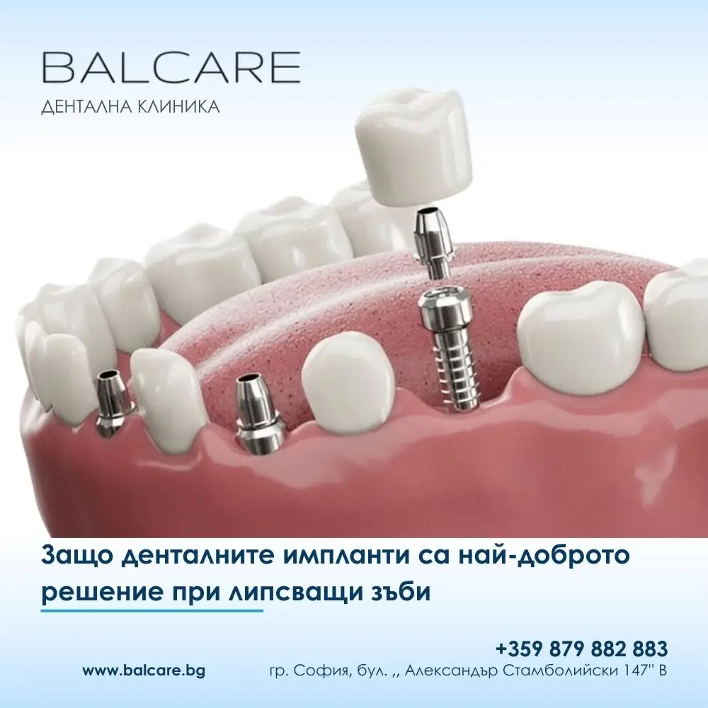
Possible Complications After Dental Implant Placement

Dental implant placement is one of the most successful and predictable procedures in modern dentistry. Implant treatment has a long-term success rate of over 98%.

However, in rare cases, complications such as implant failure, swelling, inflammatory processes, nerve involvement, damage to adjacent teeth, or injury to anatomical structures such as the maxillary sinus may occur.

These conditions usually result from a combination of factors, including the patient’s general health, the quality of the diagnostic process, the surgical technique, and the patient’s behaviour during the healing period.

Despite the high success rate, dental implant treatment is a surgical procedure and, like any operation, requires precision, careful planning, and active cooperation from the patient.

When these conditions are not met, complications may occur, some of which can compromise the final result and the overall health of the oral cavity.

Below, we discuss the most common possible complications after dental implant placement, why they occur, and how their risk can be reduced.

Dental Implant Failure – When and Why Can It Occur?

Dental implant failure is one of the less common complications in implant dentistry.

Fewer than 0.5% of patients may experience this problem, and it most commonly occurs during or shortly after the period of osseointegration.

The most common causes of implant failure are associated with a compromised immune system, chronic conditions such as diabetes or oncological disease, and infections in the implant area.

Infections may develop as a result of insufficient sterility during surgery or inadequate oral hygiene afterwards.

When the tissues surrounding the implant become inflamed, the process of osseointegration may be disrupted and the implant may remain unstable.

What Are the Symptoms of Implant Failure?

Early recognition of the symptoms is essential for preserving both the implant and the surrounding bone.

Patients most commonly report:

  • swelling that does not decrease after the first week;
  • pain;
  • redness;
  • bleeding gums.

At a more advanced stage, the following may occur:

  • unpleasant odour;
  • purulent discharge;
  • gum recession;
  • a feeling that the implant is loose.

These signs require prompt consultation with a dental professional, as timely intervention may help prevent the need to remove the implant.

A high-quality implant system, an experienced surgeon, and strict oral hygiene are among the key factors that help minimise this risk.

Swelling After Dental Implant Placement – Is It Normal?

Swelling after implant surgery is a normal response of the body.

It is usually most pronounced during the first 24 to 48 hours and gradually decreases over the following days.

The swelling results from the body’s natural inflammatory response to surgical trauma.

Blood and tissue fluids are directed towards the surgical area to support healing and protect against infection.

Cold compresses are generally recommended during the first two days, together with strict adherence to the postoperative instructions.

When healing progresses normally, the swelling subsides without additional intervention.

Inflammation Around the Implant and Peri-Implantitis

The implant itself does not become inflamed.

Inflammation affects the surrounding soft tissues and bone. When bone loss is also present, the condition is known as peri-implantitis.

One of the most common contributing factors is inadequate oral hygiene combined with a lack of regular professional cleaning.

Bacterial plaque around the implant can gradually lead to bone loss and compromise implant stability.

Prevention includes:

  • precise daily oral hygiene at home;
  • regular professional cleaning;
  • routine dental follow-up examinations.

Nerve Injury During Dental Implant Placement

Injury to the mandibular nerve is one of the most serious, although rare, complications of implant placement.

It may result in altered or reduced sensation in areas such as the lower lip, chin, or tongue.

In our clinical practice, this risk is minimised through detailed treatment planning and modern diagnostic imaging.

Three-dimensional imaging makes it possible to determine the exact distance from the planned implant site to important anatomical structures and to select the appropriate implant length and position.

The experience and precision of the implant dentist are crucial for reducing the risk of this type of complication.

Injury to the Maxillary Sinus

When teeth have been missing for many years, the bone beneath the maxillary sinus may become significantly reduced.

In such cases, there may be a risk of the implant extending towards or into the sinus cavity.

A minor sinus membrane perforation does not always lead to serious long-term problems, but the implant should be surrounded by appropriate bone support and positioned according to the treatment plan.

Rather than accepting a compromised solution, the correct approach is careful diagnostics and, when necessary, additional procedures to increase the available bone volume.

Damage to the Roots of Adjacent Teeth

An incorrectly positioned implant may damage the roots of neighbouring natural teeth.

This can occur when treatment planning is insufficient or when three-dimensional imaging is not used appropriately.

Modern implant dentistry relies on:

  • 3D imaging;
  • precise digital planning;
  • individually designed surgical guides when indicated.

These technologies allow a high degree of accuracy and help protect adjacent anatomical structures.

Frequently Asked Questions About Complications After Dental Implant Placement

What Patients Commonly Ask What You Should Know
Can the body reject a dental implant? Implant failure is uncommon – below 1% in many situations – and is most often associated with infection, impaired healing, or certain medical conditions.
How can I tell if an implant is failing? Persistent pain, swelling, bleeding, or mobility are not normal findings and require professional assessment.
Is pain normal after implant placement? Mild to moderate discomfort may be normal for several days. Severe or increasing pain requires examination.
How long is swelling normal? Swelling for approximately 2–3 days is common and should gradually decrease.
Is inflammation around the implant dangerous? It can often be treated when detected early and when good oral hygiene is maintained.
What is peri-implantitis? It is an inflammatory condition affecting the tissues and bone surrounding a dental implant.
Can an implant damage a nerve? The risk is low when appropriate diagnostics, planning, and an experienced implant clinician are involved.
What happens if the sinus is perforated? It does not always lead to a serious complication, but the situation requires appropriate assessment and management.
Can an implant damage a neighbouring tooth? The risk is low when 3D diagnostic imaging and careful treatment planning are used.
How long does an implant take to heal? Complete healing and osseointegration generally take approximately 3 to 6 months.
How can I reduce the risk of complications? Choosing an experienced clinician and maintaining excellent oral hygiene are key factors.
Does smoking affect dental implants? Smoking significantly increases the risk of healing problems and implant complications.
Can a person with diabetes receive a dental implant? Yes, implant treatment may be possible when diabetes is well controlled.
When does an implant need to be removed? Removal may be necessary when there is persistent infection or failure to achieve stable osseointegration.

How to Reduce the Risk of Complications

One of the most important steps in reducing the risk of complications is choosing a qualified and experienced implant dentist.

Professional planning and an individual approach to each patient form the foundation of successful treatment.

The patient also plays an essential role.

This includes:

  • maintaining excellent oral hygiene;
  • following postoperative instructions carefully;
  • attending regular follow-up examinations;
  • reducing smoking and alcohol consumption, particularly during the healing period.

When the clinician and patient work together as a team, a dental implant can provide a reliable, long-term solution with an excellent functional and aesthetic result.


premahvane-na-zubni-implanti-dentalna-klinika-balcare.webp
08/Jan/2026

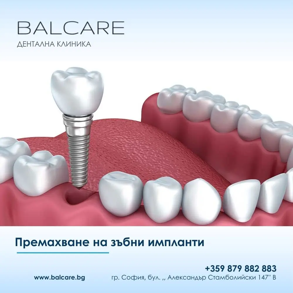
Dental Implant Removal

Dental implant removal is a complex oral surgical procedure that is performed only when there are clear medical indications.

Although dental implants are among the most reliable and long-lasting solutions for replacing missing teeth, complications can occasionally occur. These are most commonly associated with infection, failed osseointegration, inadequate treatment planning, mechanical problems, or systemic health conditions.

How Long Do Dental Implants Last?

Dental implants are designed as a long-term solution and, under favourable conditions, may remain functional for decades and sometimes for the rest of a patient’s life.

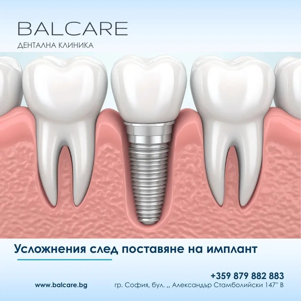
It is important to understand that an implant restoration consists of several components:

  • the implant fixture, usually made of titanium;
  • the abutment;
  • the dental crown or prosthetic restoration.

The titanium implant fixture integrates with the jawbone and has excellent biocompatibility and mechanical durability.

With good oral hygiene and regular preventive care, an implant fixture may remain stable for 25–30 years or longer without significant functional changes.

The crown is exposed directly to chewing forces and therefore generally has a shorter lifespan. Depending on the restorative material, bite, oral habits, and maintenance, it may need to be replaced after approximately 10–15 years.

This does not necessarily represent an implant complication but may simply be part of the normal long-term maintenance of an implant-supported restoration.

The experience of the implant dentist also plays an important role.

Accurate treatment planning, correct three-dimensional implant positioning, and appropriate control of chewing forces can directly affect the long-term prognosis.

Even a high-quality implant system can become compromised if it is placed incorrectly or in unsuitable bone conditions.

General health should also be considered.

Factors such as:

  • poorly controlled diabetes;
  • osteoporosis;
  • smoking;
  • chronic poor oral hygiene;

may negatively affect implant prognosis.

In patients without significant risk factors, long-term implant success rates can exceed 95%.

Are Dental Implants Safe?

Dental implant treatment is considered one of the safest and most predictable procedures in modern dentistry when it is correctly planned and performed.

However, implant placement is a surgical procedure, which means that potential risks do exist.

One possible complication is infection, which may develop around the time of surgery or during the healing period.

Factors that may contribute include:

  • inadequate oral hygiene;
  • bacterial contamination;
  • compromised healing;
  • reduced immune response.

Another critical stage is osseointegration – the biological process through which the implant becomes integrated with the surrounding bone.

If osseointegration does not occur successfully, the implant may remain mobile and functionally unstable.

This is more likely in patients with:

  • insufficient bone volume or quality;
  • active inflammation;
  • certain systemic conditions;
  • smoking-related healing impairment.

There are also anatomical risks, such as injury to a nerve or involvement of the maxillary sinus.

These complications are uncommon and can be significantly reduced through careful diagnostics and 3D imaging before implant placement.

Despite these potential risks, dental implants offer important advantages compared with conventional bridges and removable dentures.

They:

  • do not require preparation of neighbouring healthy teeth;
  • provide excellent stability;
  • help preserve the jawbone;
  • restore chewing function effectively.

For appropriately selected patients and when treatment is carefully planned, dental implants remain one of the most predictable options for replacing missing teeth.

When Might a Dental Implant Need to Be Removed?

Dental implant removal is not common, but in certain situations it may become medically necessary.

One possible reason is incorrect treatment planning or implant positioning.

Errors in the interpretation of radiographic or 3D imaging may result in an implant being placed too close to:

  • a nerve;
  • the maxillary sinus;
  • the root of a neighbouring tooth;
  • another important anatomical structure.

This may cause persistent discomfort, altered sensation, or functional problems.

Another important reason is peri-implantitis – inflammation affecting the tissues around an implant and associated with progressive loss of supporting bone.

In advanced cases, the implant may lose stability and removal may become necessary.

Systemic conditions, blood-clotting disorders, active periodontal disease, and smoking may also increase the risk of complications.

Many implant-related problems can be reduced through appropriate treatment planning, regular professional monitoring, and good home care.

Reasons for Dental Implant Removal

Reason for Removal Approximate Share of Cases When It Most Commonly Occurs Professional Explanation
Failed osseointegration 2–5% First 3–6 months The implant fails to integrate with the bone. Possible contributing factors include infection, insufficient bone quality or volume, smoking, or systemic conditions.
Early postoperative infection 1–3% Within the first 3 months Bacterial infection develops during or shortly after surgery. If it cannot be controlled, implant failure may occur.
Peri-implantitis 5–15% After 2–10 years Chronic inflammation around the implant associated with progressive bone loss. It is an important cause of late implant failure.
Incorrect planning or positioning 2–4% From 6 months to several years The implant may be placed too close to a nerve, sinus, or neighbouring tooth and may cause pain, altered sensation, or functional problems.
Insufficient bone volume 1–3% Within 1–2 years Initial stability may be achieved, but the surrounding bone may not provide adequate long-term support.
Mechanical complications 1–2% After 5–15 years Fracture or loosening may occur, particularly in patients with bruxism or an unfavourable bite. Implant fixture fracture itself is relatively uncommon.
Periodontal disease 3–6% After 3–8 years Active periodontal disease can increase the risk of inflammation and progressive bone loss around implants.
Systemic factors 1–4% At any stage Poorly controlled systemic conditions may interfere with healing and long-term maintenance.
Lack of maintenance and preventive care Up to 20%* After 5+ years *This is not necessarily a direct cause but may contribute significantly to many late complications.

In many cases requiring implant removal, the problem is not caused by the implant material itself but by a combination of factors such as:

  • inadequate oral hygiene;
  • lack of regular preventive examinations;
  • smoking;
  • untreated periodontal disease.

Early implant failures occurring within the first six months are relatively uncommon and are usually associated with biological or surgical factors.

Late failures, which occur after several years, are more frequently associated with maintenance, inflammation, and biomechanical loading.

What Does Clinical Practice Show?

  • Overall long-term implant success rates are approximately 94–97% at 10-year follow-up.
  • Regular maintenance appointments may substantially reduce the risk of late peri-implant complications.
  • Many implant-related problems can be managed more successfully when they are identified at an early stage.

How Is a Dental Implant Removed?

Dental implant removal is a carefully planned procedure whose main objective is to remove the implant while preserving as much surrounding bone as possible.

The process begins with a detailed clinical and radiographic examination.

The implant dentist must determine:

  • why the implant needs to be removed;
  • whether it is mobile;
  • how strongly it is integrated with the bone;
  • the amount of remaining supporting bone;
  • the position of nearby anatomical structures.

This assessment determines the most appropriate removal technique.

Removal of a Mobile or Partially Integrated Implant

When an implant is mobile or only partially integrated, a minimally invasive approach may be possible.

Specialised implant removal instruments can be used to apply controlled reverse torque, allowing the implant to be carefully unscrewed from the bone.

This technique can help preserve a greater amount of the surrounding bone.

Removal of a Fully Osseointegrated Implant

If the implant is completely integrated with the bone, a more complex surgical approach may be necessary.

The procedure may include:

  1. administration of local anaesthesia;
  2. a small incision in the gum;
  3. exposure of the implant;
  4. controlled removal of a limited amount of surrounding bone;
  5. careful extraction of the implant;
  6. cleaning and assessment of the surgical site.

The exact technique depends on the implant position, degree of integration, and condition of the surrounding tissues.

What Happens After Dental Implant Removal?

After implant removal, the area is carefully assessed.

Postoperative care may include:

  • appropriate oral hygiene;
  • prescribed medication when indicated;
  • pain control;
  • regular follow-up appointments;
  • monitoring of the healing process.

If significant bone loss is present, bone regeneration or bone grafting may be recommended.

The aim is to restore sufficient bone volume and create favourable conditions for future treatment.

In selected cases, another dental implant may later be placed after the bone and soft tissues have healed adequately.

Can a New Implant Be Placed After Removal?

In many cases, yes.

Whether another implant can be placed depends on:

  • the reason the original implant failed;
  • the amount of remaining bone;
  • the presence or absence of infection;
  • the condition of the gum tissue;
  • the patient’s general health;
  • the position required for the new implant.

Sometimes a new implant can be placed immediately after removal, but in other cases the area must first heal or undergo bone regeneration.

The treatment plan is always determined individually.

How Can the Risk of Dental Implant Removal Be Reduced?

Risk Factor What Increases the Risk How the Risk Can Be Reduced Expected Benefit
Poor oral hygiene Plaque and bacterial accumulation around the implant Brush twice daily and use interdental brushes, floss, or other recommended cleaning aids Reduces the risk of peri-implant inflammation
Lack of preventive examinations Inflammation and bone loss may remain undetected Regular follow-up and professional cleaning Allows complications to be identified earlier
Smoking Reduced blood supply and delayed healing Stop or significantly reduce smoking, especially during healing Improves healing and implant prognosis
Poorly controlled diabetes Delayed healing and increased susceptibility to infection Maintain appropriate blood glucose control in coordination with the treating physician Supports more predictable healing
Periodontal disease Ongoing inflammation and bacterial burden Treat periodontal disease before and after implant placement Improves long-term stability
Bruxism Excessive mechanical loading Individual night guard when clinically indicated May reduce the risk of loosening and mechanical complications
Incorrect bite Uneven distribution of chewing forces Occlusal analysis and adjustment where necessary Helps reduce excessive implant loading
Insufficient bone volume Reduced primary stability and support Bone augmentation before or during implant placement when indicated Improves the conditions for implant stability
Failure to follow postoperative instructions Increased risk of infection and impaired healing Follow all postoperative recommendations carefully Helps reduce early complications
Lack of patient education Incorrect home-care habits Clear instructions on implant maintenance Supports long-term implant health

A dental implant does not usually “fail on its own”.

Complications are generally multifactorial and may result from a combination of biological, mechanical, behavioural, and systemic factors.

When the patient and implant team work together through good home care, regular professional maintenance, and early management of problems, the risk of implant loss can be significantly reduced.

Dental Implant Removal Is Sometimes Part of Responsible Treatment

Removing a dental implant should not automatically be viewed as a treatment failure.

In certain situations, it is the most appropriate medical decision to protect the surrounding bone, soft tissues, neighbouring teeth, or the patient’s overall health.

With timely diagnosis, an experienced implant dentist, and appropriate postoperative care, implant removal can usually be performed safely while preserving as much bone as possible.

When conditions allow, bone regeneration and future implant treatment can often restore the area again and provide a stable long-term result.


10-prichini-zashto-dentalnite-implanti-sa-nay-dobroto-reshenie-pri-lipsvashti-zabi-balcare.webp
02/Jan/2026

10 Reasons Why Dental Implants Are the Best Solution for Missing Teeth

Losing a tooth is never only an aesthetic issue. It affects the way you eat, speak, smile, and even how confident you feel.

In our clinical practice, we see every day how a single missing tooth can gradually lead to more serious functional and oral health changes.

Modern dentistry, however, offers a solution that comes remarkably close to a natural tooth – the dental implant.

Below, we explain clearly and professionally why dental implants are considered the gold standard for replacing missing teeth and why, in many cases, they offer important advantages over alternative solutions.

1. Dental Implants Provide a Long-Term and Stable Solution

A dental implant is designed to become a permanent part of your mouth.

Once the implant has integrated with the surrounding bone, it functions as an artificial tooth root and provides excellent stability.

With good oral hygiene and regular preventive examinations, dental implants can last for decades and, in many cases, for the rest of the patient’s life.

This makes them significantly more reliable in the long term than removable dentures or dental bridges, which may require replacement over time.

2. The Closest Feeling to a Natural Tooth

Dental implants do not only look like natural teeth – they can also feel remarkably similar.

Patients often report that after an adaptation period, they forget which tooth has been replaced by an implant.

The crown placed on the implant is individually designed according to:

  • shape;
  • colour;
  • size;
  • the surrounding natural teeth.

This allows the restoration to blend naturally into the smile and restore normal function during eating and speaking.

3. Preservation of Healthy Adjacent Teeth

One of the major advantages of dental implants is that they do not require preparation of the neighbouring teeth.

With a conventional dental bridge, healthy adjacent teeth usually need to be reduced so that they can support the restoration.

A dental implant is an independent structure.

This means that neighbouring natural teeth can remain intact, which is an important advantage for long-term oral health.

4. Greater Freedom When Eating

Dental implants allow patients to eat with confidence.

They provide excellent stability and chewing function that can closely resemble that of natural teeth.

Foods such as:

  • meat;
  • nuts;
  • fresh fruit;
  • firmer foods;

can generally be eaten with considerably greater confidence than with an unstable removable prosthesis.

Restoring effective chewing not only improves quality of life but may also support digestion by allowing food to be properly broken down before swallowing.

5. Clear and Confident Speech

Missing teeth may affect pronunciation and speech.

Certain sounds may become less clear, which can make patients feel self-conscious when communicating.

Dental implants restore the anatomy of the missing tooth area and can support clear and confident speech without the fear of a removable prosthesis shifting or becoming displaced.

6. Helping Preserve the Jawbone

After a tooth is lost, the bone in that area gradually begins to resorb.

This is a natural biological process that, over time, may contribute to changes in facial contours and a more aged appearance.

A dental implant transfers functional forces to the surrounding bone in a way that is similar to a natural tooth root.

This helps maintain bone volume in the area and supports the natural contours of the jaw and face.

7. Straightforward Maintenance and Oral Hygiene

Dental implants do not require complicated daily maintenance.

They are cared for in a way similar to natural teeth, including:

  • regular brushing;
  • interdental cleaning or flossing;
  • professional preventive care.

Unlike removable dentures, they do not need to be removed, fixed in place with adhesive, or soaked in special solutions.

This makes everyday maintenance considerably more convenient for many patients.

8. A Very High Success Rate

Modern clinical studies report dental implant success rates of more than 95%.

This makes implant treatment one of the most predictable procedures in modern dentistry.

Success depends on several important factors, including:

  • accurate treatment planning;
  • the experience of the implant dentist;
  • appropriate surgical technique;
  • good oral hygiene;
  • regular follow-up care.

9. An Individual and Precise Treatment Approach

There is no universal implant solution that is suitable for every patient.

Each person has different:

  • bone anatomy;
  • bite;
  • soft tissues;
  • general health;
  • aesthetic requirements.

Implant treatment is therefore individually planned from the selection and positioning of the implant to the design of the final crown.

This allows the clinician to optimise both the functional and aesthetic result.

10. Greater Confidence and Improved Quality of Life

A complete and healthy-looking smile can change the way people feel about themselves.

Many patients become more confident, more comfortable in social situations, and more relaxed in their everyday lives.

Dental implants do more than simply replace a missing tooth.

They can restore comfort, confidence, function, and quality of life.

Dental Implants Compared with Other Tooth-Replacement Options

Dental Implants vs Removable Dentures

Removable dentures may move during eating or speaking, cause discomfort, and require a period of adaptation.

Dental implants are fixed and stable and do not create the same sensation of a removable foreign object in the mouth.

Dental Implants vs Dental Bridges

Dental bridges generally require preparation of neighbouring teeth and do not prevent bone loss in the area of the missing tooth.

Dental implants can help preserve both the surrounding bone and the healthy adjacent teeth.

Comparison of Dental Implants, Bridges and Removable Dentures

Criterion Dental Implants Dental Bridges Removable Dentures
Durability 20+ years, often lifelong 7–12 years 5–8 years
Stability Fixed in the bone and highly stable Fixed, but dependent on adjacent teeth May move during function
Effect on neighbouring teeth No preparation required Healthy teeth generally need preparation No direct preparation, but they may be subjected to additional loading
Bone preservation Yes – helps stimulate and maintain the bone No No
Comfort Very high, close to the feeling of a natural tooth Good, but not completely natural May cause discomfort
Aesthetics Excellent, highly natural appearance Good Generally lower
Maintenance Similar to natural teeth Requires careful cleaning around and beneath the bridge Must be removed and cleaned
Chewing efficiency 90–100% 60–80% 30–50%
Confidence and sense of security Very high Moderate Often lower

How Should You Make Your Decision?

The comparison clearly demonstrates why dental implants are widely considered a gold-standard option for replacing missing teeth.

They do not simply restore the appearance of the smile. They also provide an important biological benefit that other options cannot offer to the same extent – supporting preservation of the jawbone.

With dental bridges, the main compromise is the preparation of healthy neighbouring teeth.

Over the long term, this may lead to the need for additional dental treatment and higher costs.

Although bridges may initially appear to be a faster solution, they do not stop bone resorption beneath the missing tooth.

Removable dentures are often selected because of financial considerations, but they may require ongoing adaptation.

Patients may report difficulties with:

  • eating;
  • speaking;
  • social situations;
  • stability and comfort.

These factors can directly affect quality of life.

Which Option Is Most Suitable for You?

The most appropriate treatment always depends on the individual patient.

Important factors include:

  • available bone volume;
  • general health;
  • oral hygiene;
  • lifestyle habits;
  • bite and functional loading;
  • individual expectations.

However, when implant treatment is clinically possible, dental implants can provide an excellent combination of stability, aesthetics, function, and long-term reliability.

From a clinical perspective, investing in a dental implant may also prove more economical in the long term because it can reduce the need for frequent adjustments, replacements, and additional interventions.

Professional Advice from an Implant Dentist

If you are considering several tooth-replacement options, do not make the decision based only on price or speed of treatment.

The more important question is how the chosen solution will affect your oral health 10, 15, or 20 years from now.

A consultation with a dental implant specialist can provide you with a clear, personalised treatment plan and a realistic understanding of the most appropriate option for your individual case.

If you are considering treatment, professional consultation is the first and most important step.

Accurate diagnosis and an individual approach are the foundation of a successful and long-lasting result.

A smile can change your life – and often, it begins with the right decision.


payment-cards-dentalna-klinika

Copyright by BalCare2026. All rights reserved.

Call Now Button